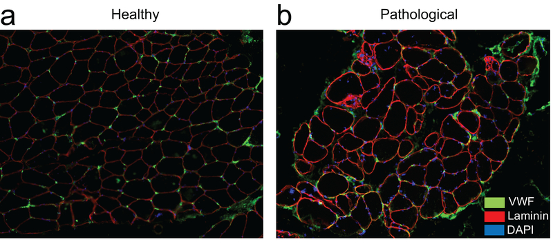
Figure 3.
Representative immunohistochemical cross-sections of human paraspinal muscle from normal (A) and pathological (B) human paraspinal muscle illustrating the distribution and location of capillaries (green) in-between myofibers (outlined in red). Myofiber nuclei are stained in blue. Reduced capillary density and clustering as well as heterogeneous myofiber size and distribution can be observed in the pathological sample.

