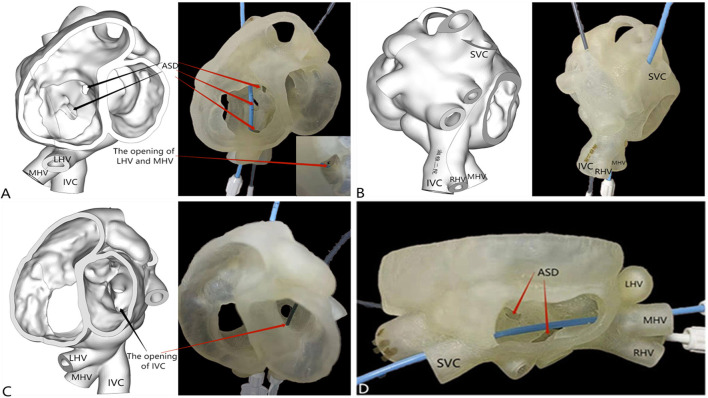
Figure 2.
3D reconstruction and 3D printing. (A) Three large atrial septal defects (one of fossa ovalis type [diameter of 6 mm], one near the superior vena cava [diameter of 15 mm], and one near the inferior vena cava [diameter of 8 mm]); the left hepatic vein joined the middle hepatic vein, and the opening was located in the right atrium. (B,C) The right hepatic vein drained into the inferior vena cava, and the opening of the inferior vena cava was located in the left atrium. (D) Two atrial septal defects were visible from the right atrium incision level.

