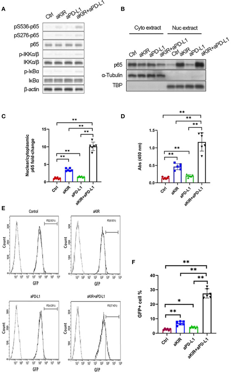
Figure 3.

Lirilumab and Avelumab Co-Engagement Disinhibits NF-κB. (A) IL-2-primed NK cells were mixed with autologous target cells and stimulated with lirilumab (aKIR) and/or avelumab (aPD-L1) by receptor crosslinking for 4 h. Whole cell lysates were immunoblotted for phospho-S536 p65, phospho-S276 p65, total p65, phospho-S176/180 IKKα/β, total IKKα/β, phospho-IκBα, total IκBα, and β-actin. (B, C) IL-2-primed NK cells were mixed with autologous target cells and stimulated with plate-immobilized aKIR and/or aPD-L1 for 1 h. Cytoplasmic and nuclear extracts were immunoblotted with anti-p65 antibody. The ratios of normalized nuclear p65:normalized cytoplasmic p65 are reported. (D) Nuclear extracts collected as in (B) were added to a 96-well plate immobilized with the consensus NF-kB-binding sequence. p65 bound to the oligonucleotide was measured by colorimetric assay. (E, F) IL-2-primed NK cells transduced with a κB-GFP reporter construct were mixed with autologous target cells and stimulated with plate-immobilized aKIR and/or aPD-L1 for 6 h. GFP expression in NK-κB-GFP cells was analyzed by flow cytometry. *P < 0.05, **P < 0.01 [one-way ANOVA]. n=3 biological replicates×3 technical replicates.
