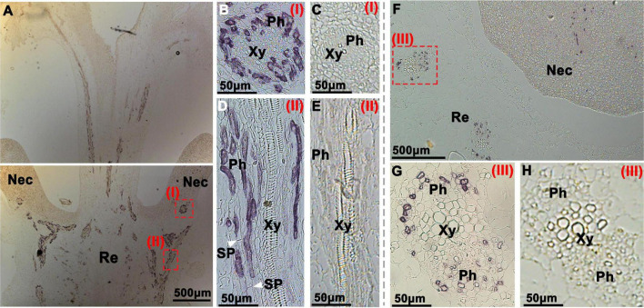
FIGURE 4.
Immunohistochemical localization of CsSWEET7a in female cucumber flowers. Longitudinal (A–E) and transverse sections (F–H) of female flowers with AP as the second antibody. (B,D,G) The close-up of the boxes in panels (A,F), respectively. The white arrows in panel (D) indicate the sieve plate. (C,E,H) Sections were incubated with pre-immune serum as a control. Images in panels (G,H) were rotated 90°clockwise. The Roman numerals in red at the top-right corner (B–E,G,H) correspond to those fields in panels (A,F). Nec, nectary; Re, receptacle; Ph, phloem; Xy, xylem; SP, sieve plate.

