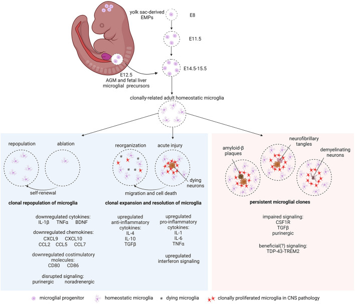
Figure 1.
Microglial clonality in development, health, and disease. Yolk sac EMPs enter the mouse neuroepithelium from E8 to E11.5 where they clonally expand and colonize the brain niche. An additional influx of Hoxb8+ microglial precursors from the AGM and fetal liver at E12.5 invade and populate the brain between E14.5 and E15.5 to make up the rest of the postnatal resident microglial population. Microglia clonally repopulate after ablation via self-renewal (left panel). In acute injury, microglia respond by microgliosis (clonal proliferation) at the site of damage (center panel). With clinical recovery, clones of microglia are reorganized by microglial cell migration and cell death (center panel). In chronic neurodegeneration such as AD, PD and MS, microglial clones persist around Aβ plaques, neurofibrillary tangles (tauopathy), and demyelinating neurons (right panel). Altered gene regulation and signaling pathways mentioned in the text are indicated. Created with BioRender.com.

