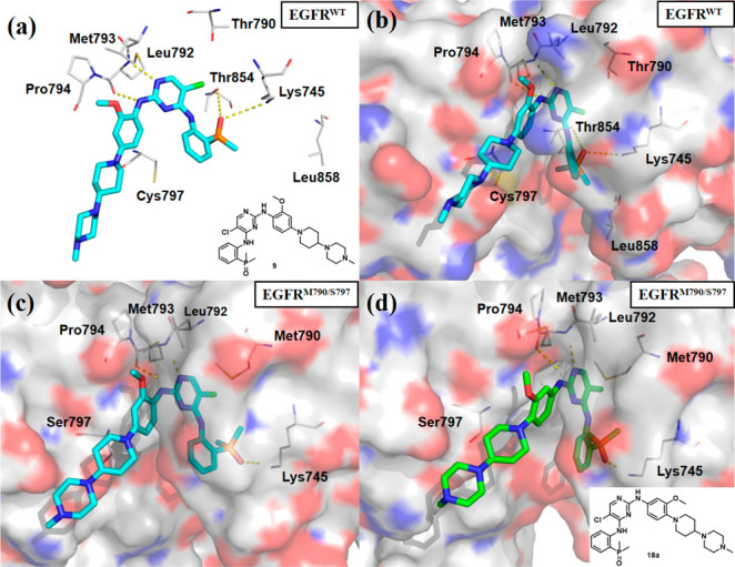
Figure 2.

X-ray crystal structure of brigatinib (9) with EGFRThr790/Cys797(EGFRWT) kinase domain (PDB 7AEM) shown in cyan and gray in stick and line representation (a) or in surface representation (b). Preliminary computational docking complex of 9 (c) and 18a (d) with EGFRMet790/Ser797 (PDB 5ZTO). Hydrogen bonds are indicated by yellow hatched lines to key amino acids.
