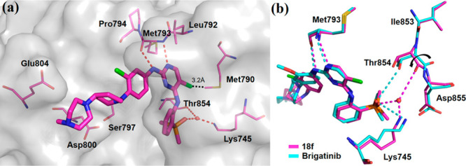
Figure 3.

(a) X-ray crystal structure of EGFRT790M/C797S in complex with 18f (PDB 7ER2). Key residues and 18f are shown in magenta by the element in lines and in sticks, respectively. Hydrogen bond interactions are showed as red dotted lines, and the water molecule is indicated by a red dot. (b) Superposition of 18f (PDB 7ER2; compound and protein shown as magenta sticks and lines, respectively) and brigatinib complex (PDB 7AEM; compound and protein shown as cyan sticks and lines, respectively).
