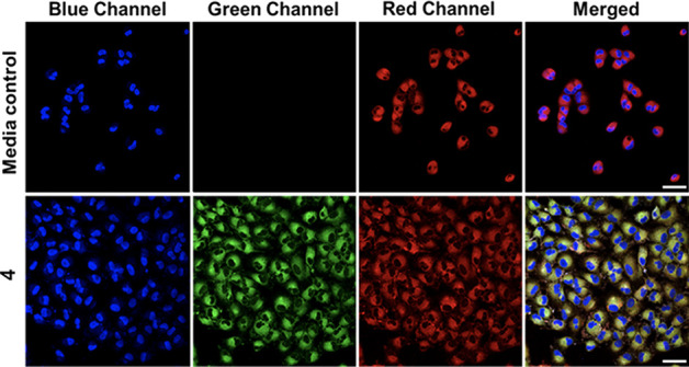
Figure 4.

Co-localization of 4 with ER-Tracker Red in A549 cells. Cells were treated with vehicle control (0.4% DMSO) (row 1), or 4 (25 μM, row 2) for 1 h, then counterstained with Hoechst 33342 and ER-Tracker Red. These images depict the Hoechst nuclear stain (blue channel), 4 (green channel), ER-Tracker Red (red channel), and a merge of the three channels. Scale bar = 50 μm.
