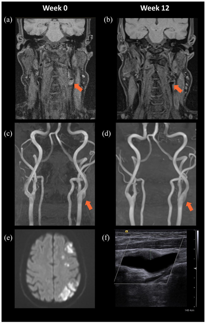
Figure 2.
(a–e) Confirmation of left ICA dissection in MRI and follow-up images after 12 weeks. MRI with T1-weighted, fat-saturated images (a–b) and time-of-flight angiography (c–d) showing suspicious ICA dissection in week 0 (a, c) and improvement at follow up in week 12 (b, d). (e) Diffusion-weighted imaging at week 0 shows a diffusion restricted pattern in the territory of the left medial cerebral artery matching haemodynamic infarction. (f) Follow-up neurovascular ultrasound at 12 weeks shows receding vessel wall hematoma (dotted green line: 4.5 mm).
ICA, internal carotid artery; MRI, magnetic resonance imaging.

