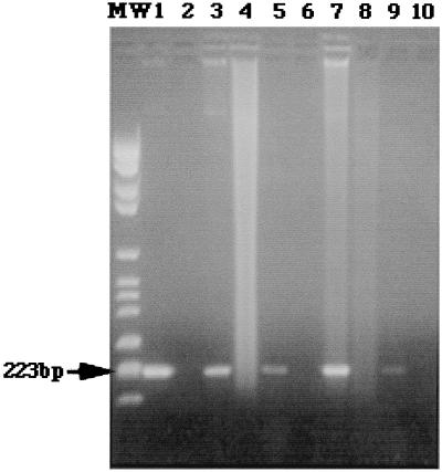
FIG. 1.
Agarose gel electrophoresis and ethidium bromide staining. Lane MW, DNA ladder (223 bp); lane 1, positive control (B. abortus B-19 DNA); lane 2, a sample to which no DNA was added; lane 3, synovial fluid from a brucellosis patient with knee arthritis; lane 4, synovial fluid from a patient with knee arthritis due to S. aureus; lane 5, urine sample from a patient with orchiepididymitis due to B. melitensis; lane 6, urine sample from a patient with E. coli pyelonephritis; lane 7, sample of pus from a liver abscess due to B. melitensis; lane 8, sample of pus from a liver abscess due to E. coli; lane 9, CSF from a brucellosis patient with meningitis; lane 10, CSF from a patient with meningitis due to M. tuberculosis. The photocomposition of the figure was obtained from the original Polaroid films with a ScanJet IIcx scanner (Hewlett-Packard, Corvallis, Oreg.) After the initial image was scanned and saved as a tagged image file format file, the file was opened in Adobe Photoshop (Adobe System, Inc., Seattle, Wash.).

