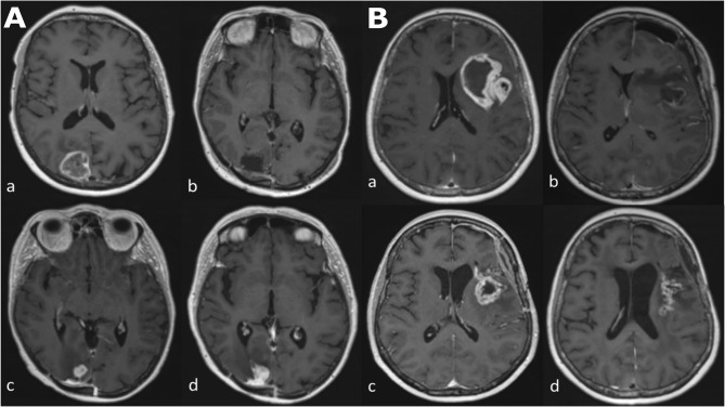
Figure 1.
Longitudinal series of MRI images in two patients (A, B) with glioblastoma, IDH-wildtype. All images are axial T1-weighted after contrast administration. Images (Aa–Ad) demonstrate tumor progression. (Aa) Pre-operative MRI of a glioblastoma in the occipital lobe. (Ab) Post-operative MRI five days after resection; there is no contrast enhancement therefore no identifiable residual tumor. (Ac) The patient underwent a standard care regimen of radiotherapy and temozolomide. A new enhancing lesion at the inferior margin of the post-operative cavity was identified on MRI at three months after radiotherapy completion. (Ad) The enhancing lesion continued to increase in size three months later and was confirmed to represent tumor recurrence after repeat surgery. Images (Ba–Bd) demonstrate pseudoprogression. (Ba) Pre-operative MRI of a glioblastoma in the insula lobe. (Bb) Post-operative MRI at 24 hours after surgery; post-operative blood products are present but there is no contrast enhancement therefore no identifiable residual tumor. (Bc) The patient underwent a standard care regimen of radiotherapy and temozolomide. A new rim-enhancing lesion was present on MRI at five months after radiotherapy completion. (Bd) Follow-up MRI at monthly intervals showed a gradual reduction in the size of the rim-enhancing lesion without any change in the standard care regimen of radiotherapy and temozolomide or corticosteroid use. The image shown here is the MRI four months later.

