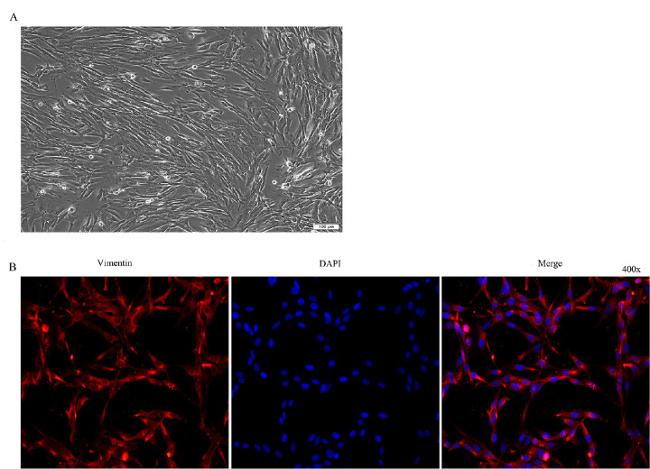
Figure 1.
Culture and identification of HTFs. (A) Morphological observation of primary cultured cells of HTFs (100 × inverted microscope). (B) Immunofluorescence identification (400 × inverted fluorescence microscope). Note: Fluorescence (Cy3) labeled goat anti-rabbit IgG labeling (red), DAPI staining (blue).

