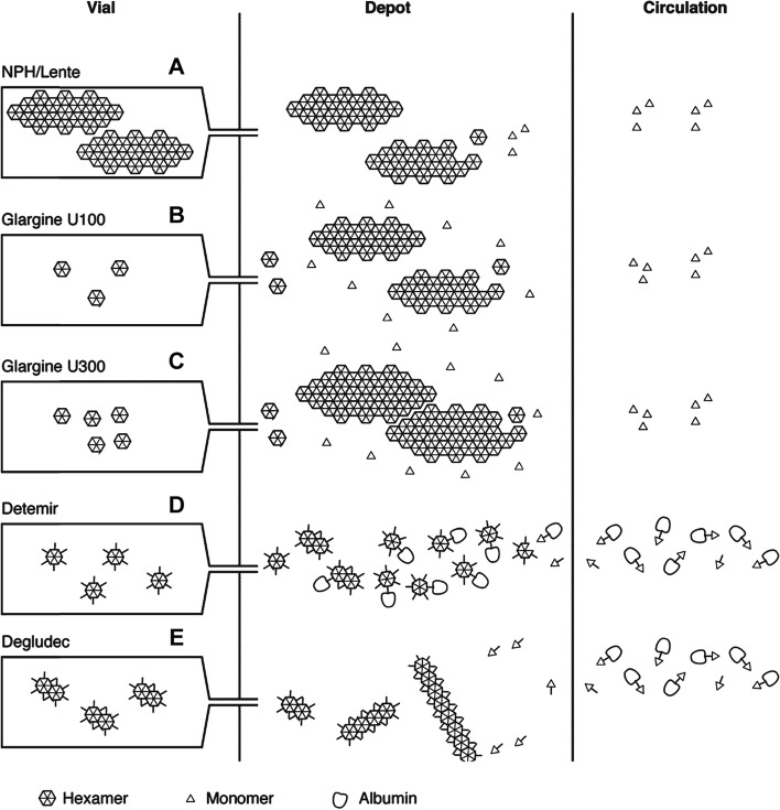
Fig. 4.
Summary of the mechanism of protraction of insulin action. Insulins vary in the size and type of crystal formed at the site of injection, and the ability to interact with albumin. NPH insulin (A) is injected as a pre‐formed protein–insulin conglomerate. Insulin glargine (100 U/mL) (B) forms crystals when the pH increases following injection. Insulin glargine 300 U/mL (C) also precipitates at physiological pH but these precipitates are more compact compared with the 100 U/mL preparation, reducing the surface area for absorption. Insulin detemir (D) hexamers self-associate at the injection site into dihexamers, thereby slowing absorption, and reversibly bind to albumin both in subcutaneous tissues and in circulation. Insulin degludec (E) has similar properties, but further protraction of absorption is achieved by multihexamer chain formation at the site of injection. Subsequent dissociation of zinc causes the terminal hexamers to break down. Modified from Heise and Mathieu, 2017 [25]

