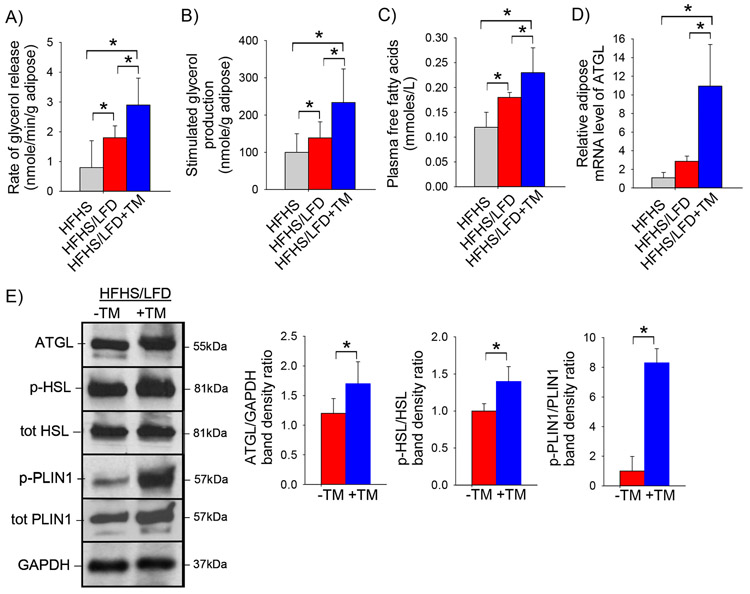
Figure 3: PAI-1 inhibition promotes adipose tissue lipolysis.
A) Rate of glycerol release from adipose tissue (nmole/min/g), B) isoproterenol-stimulated adipose tissue glycerol production (nmole/g adipose), C) plasma total free fatty acid concentration (nmole/L), D) relative adipose tissue mRNA expression of Atgl, and E) representative western blot of ATGL, phosphorylated and total HSL and phosphorylated and total PLIN1 protein level in pooled adipose tissue protein samples with corresponding densitometric analysis in C57BL/6J mice fed a high-fat, high-sugar diet for 8 weeks (HFHS) then either a low-fat diet (LFD) or low-fat diet supplemented with TM5441 (LFD+TM) for an additional 2 weeks. Values are expressed as mean (n=6) ± SD, * p<0.05.

