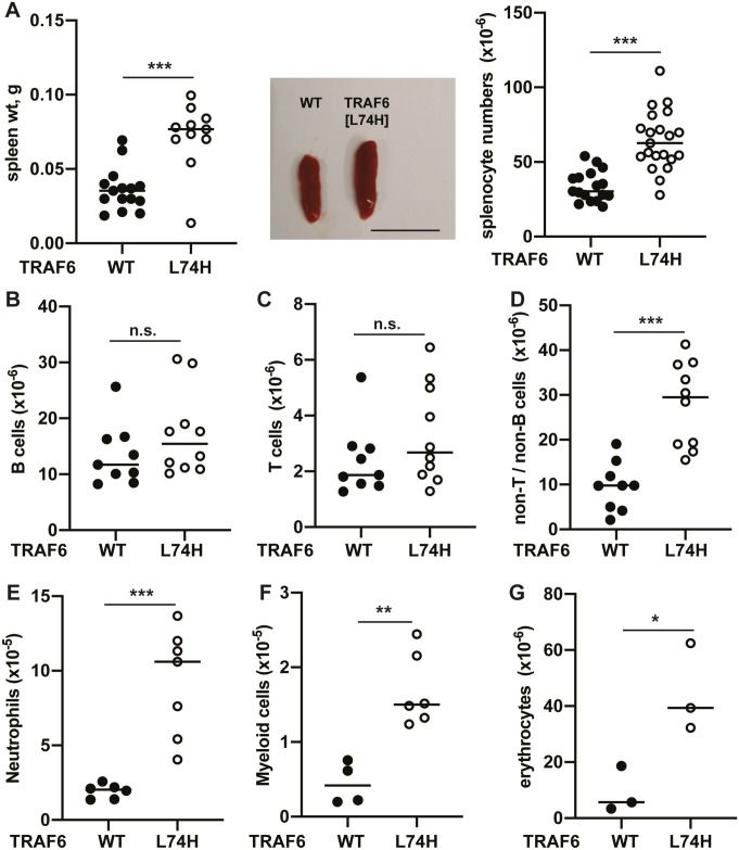
Fig 2. Characterization of splenic immune cell populations in TRAF6[L74H] mice.
(A) Spleen weight (left panel) and representative images of spleen enlargement (middle panel) and total splenocyte numbers of 16 days old WT (n = 15–17) and TRAF6[L74H] (n = 11–21) mice, scale bar = 1 cm. (B-D) Immune cell populations in the spleen of 16 days old WT and TRAF6[L74H] mice were analyzed by flow cytometry. Total numbers of (B) B-cells, (C), T-cells, (D) and non-T/B cells in spleens of WT (n = 9) and TRAF6[L74H] (n = 10) mice are shown. (E-G), as in B-D, except that (E) neutrophils from WT (n = 6) and TRAF6[L74H] (n = 7) mice (F) Gr-1-CD11b+ myeloid cells from WT (n = 4) and TRAF6[L74H] (n = 6) mice or (G) erythrocytes WT (n = 3) and TRAF6[L74H] (n = 3) mice were measured. Symbols represent individual biological replicates. Statistical significance between the two genotypes was calculated using the unpaired t-test with Welch’s correction or the Mann-Whitney test. * denotes p<0.05, ** denotes p<0.01, *** denotes p<0.001 and n.s. denotes not significant difference. Individual values, descriptive statistics and results from the statistical analysis are provided in S2 File.

