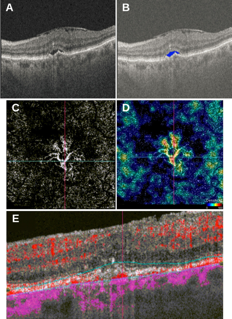
Fig 3.

(A) SS-OCT volume scan of 87-year-old female patient with type I CNV, SRF and epiretinal membrane (ERM); (B) fluid segmentation using a convolutional neural network (CNN) highlights SRF in blue; (C) corresponding OCT-A scan depicting segmented CNVM from outer retina (OR) slab; (D) density flow highlighting areas of increased flow; (E) corresponding flow B-scan from single horizontal B-scan through center of CNVM.
