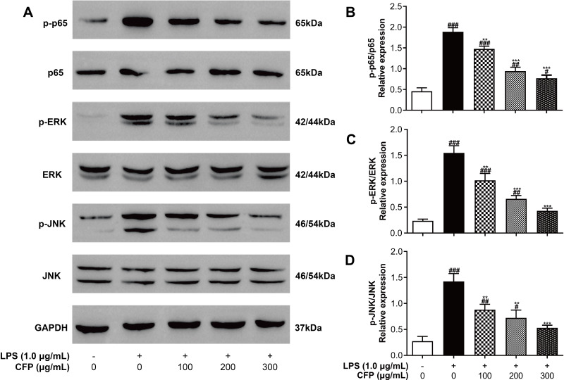
Figure 6.
Effects of CFP on the LPS-induced activation of NF-κB and MAPK pathways. (A) Represents the Western immunoblots for p-p65, p65, p-ERK, ERK, p-JNK, JNK and GAPDH. After inflammatory stimulation, relative expression of p-p65/p65 (B), p-ERK/ERK (C) and p-JNK/JNK (D) were tested by Western blotting in THP-1 macrophages. Data represent the average of the three replicates. #P < 0.05, ##P < 0.01 and ###P < 0.001 vs control group; **P < 0.01 and ***P < 0.001 vs LPS group.

