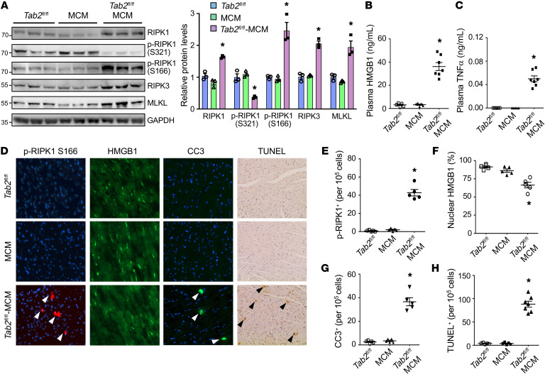
Figure 2. Loss of TAB2 promotes myocardial apoptosis and necroptosis.
(A) Western blot (left) and quantification (right) of the indicated proteins normalized to GAPDH in cardiac extracts from Tab2fl/fl, MCM, and Tab2fl/fl-MCM mice 2 weeks after tamoxifen treatment. (B and C) Plasma HMGB1 and TNF-α levels from mice indicated in A. *P < 0.05 versus Tab2fl/fl or MCM. n = 5–7. (D) Immunofluorescence staining with the indicated antibodies as well as TUNEL assay in cardiac sections from mice of the indicated genotypes. (E–H) Quantification of phospho-RIPK1 Ser166, nuclear HMGB1, cleaved caspase-3 (CC3), and TUNEL-positive cells. *P < 0.05 versus Tab2fl/fl or MCM. n = 5–7. Statistical analysis was performed using 1-way ANOVA with Tukey’s post hoc test.

