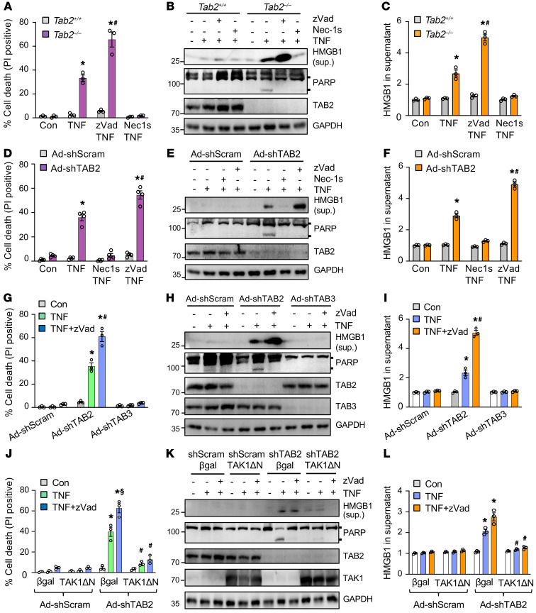
Figure 4. TAB2 but not TAB3 is a key regulator of apoptotic and necroptotic cell death in cardiomyocytes.
(A) Cell death assessed by propidium iodide (PI) staining of Tab2+/+ and Tab2–/– MEFs treated with 10 ng/ mL TNF-α or vehicle control for 6 hours in the presence or absence of necrostatin-1s (Nec-1s; RIPK1 inhibitor) or zVad-fmk (zVad; pan-caspase inhibitor). *P < 0.01 versus control; #P < 0.05 versus Tab2–/– TNF. n = 3. (B) Western blotting for the indicated proteins. Sup., culture supernatant. (C) HMGB1 in cell culture supernatant. *P < 0.05 versus control; #P< 0.05 versus Tab2–/– TNF. n = 3. (D) Cell death in neonatal cardiomyocytes infected with an adenovirus encoding TAB2 shRNA (shTAB2) or a scrambled sequence (shScram), and treated as indicated. *P < 0.01 versus control; #P < 0.05 versus shTAB2 TNF. n = 4. (E) Western blotting for the indicated proteins. (F) HMGB1 in cell culture supernatant. *P < 0.05 versus control; #P < 0.05 versus shTAB2 TNF. n = 3. (G) Cell death in neonatal cardiomyocytes treated as indicated. *P < 0.01 versus control; #P < 0.05 versus shTAB2 TNF. n = 3. (H) Western blotting for the indicated proteins. (I) HMGB1 in supernatant. *P < 0.05 versus control; #P < 0.05 versus shTAB2 TNF. n = 3. (J) Cell death in neonatal cardiomyocytes treated as indicated. TAKΔN indicates the constitutively active TAK1 mutant. *P < 0.01 versus control; #P < 0.05 versus β-gal in the corresponding group; §P < 0.05 versus Ad-shTAB2 β-gal TNF. n = 3. (K) Western blotting for the indicated proteins. (L) HMGB1 in supernatant. *P < 0.05 versus control; #P < 0.05 versus β-gal in the corresponding group. n = 3. Statistical analysis was performed using 2-way ANOVA with Tukey’s multiple-comparison test.

