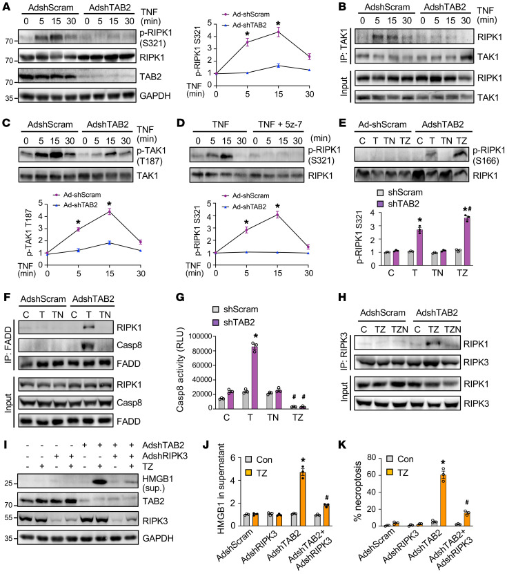
Figure 5. Loss of TAB2 promotes RIPK1-dependent apoptosis and necroptotic signaling.
(A) Western blotting for the indicated proteins in neonatal cardiomyocytes treated as indicated. *P < 0.05 versus Ad-shTAB2. n = 3. (B) Western blotting for the indicated proteins after IP with anti-TAK1 from neonatal cardiomyocytes treated as indicated. (C) Western blotting and quantification of phospho-TAK1 Thr187 in neonatal cardiomyocytes treated as indicated. *P < 0.05 versus Ad-shTAB2. n = 3. (D) Western blotting and quantification of phospho-RIPK1 Ser321 in neonatal cardiomyocytes treated as indicated. 5z-7, 5z-7-oxozeaenol. *P < 0.05 versus Ad-shTAB2. n = 3. (E) Western blotting and quantification of phospho-RIPK1 Ser166 in neonatal cardiomyocytes infected with AdshScram or AdshTAB2 followed by treatment with vehicle control (C) or TNF-α (T) in the presence or absence of necrostatin-1s (N) or zVad-fmk (Z) for 2 hours. *P < 0.05 versus control; #P < 0.05 versus shTAB2 T. n = 3. (F) Western blotting for the indicated proteins after IP with anti-FADD from neonatal cardiomyocytes treated as indicated. (G) Caspase-8 activity in neonatal cardiomyocytes. *P < 0.01 versus control; #P < 0.05 versus shTAB2 TNF. n = 3. (H) Western blotting for the indicated proteins after IP with an anti-RIPK3 antibody from neonatal cardiomyocytes treated as indicated. (I) Western blotting for the indicated proteins in neonatal cardiomyocytes infected with the indicated adenoviral vectors for 24 hours followed by treatment with TNF-α and zVad-fmk (TZ) for 6 hours. (J) Quantification of HMGB1 in cell culture supernatant. *P < 0.05 versus control; #P < 0.05 versus Ad-shTAB2 TZ. n = 3. (K) Necroptosis in cells treated as indicated in I. *P < 0.01 versus control; #P < 0.05 versus Ad-shTAB2 TZ. n = 3. Statistical analysis was performed using 2-way ANOVA with Tukey’s multiple-comparison test.

