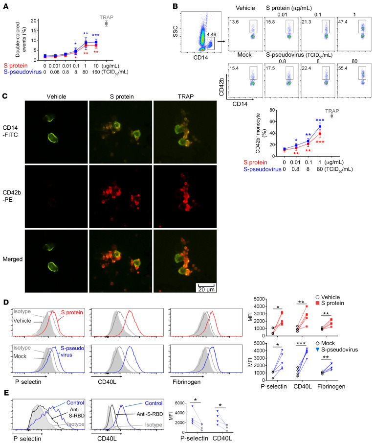
Figure 1. SARS-CoV-2–induced platelet-monocyte aggregation and P selectin/CD40L expression on platelets by spike protein.
(A) PKH26- or CFSE-labeled platelets from healthy donors (n = 5) were mixed at 1:1 and incubated with increasing concentrations of spike protein or S-pseudovirus, and aggregates were detected by flow cytometry. Double-colored events indicated the platelet aggregation, and TRAP was used as the positive control. (B) Peripheral blood from healthy donors (n = 5) was stimulated by spike protein or S-pseudovirus at indicated concentration and analyzed by flow cytometry. Platelet-monocyte aggregation was evaluated using the percentage of CD42b+/CD14+ cells. (C) Platelet-monocyte aggregation was visualized by fluorescence microscopy; scale bar: 20 μm. (D) Purified platelets from healthy donors (n = 5) were incubated with spike protein or S-pseudovirus. P selectin, CD40L expression, and fibrinogen binding were shown by histogram and MFI. (E) Purified platelets (n = 4) were pretreated with isotype antibody (indicated as control) or anti-spike RBD before incubation with S-pseudovirus; P selectin and CD40L expression are shown. Mean with SD and P value by paired Student’s t test are displayed. *P < 0.05; **P < 0.01; ***P < 0.001.

