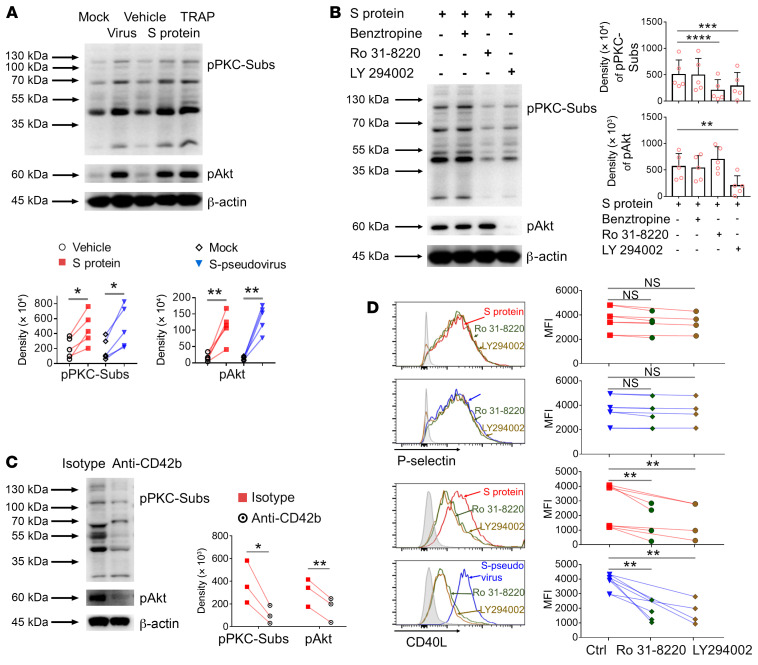
Figure 3. SARS-CoV-2 spike protein activated platelets via 2 distinct signaling pathways.
(A) Purified platelets from healthy donors (n = 5) were incubated with spike protein or S-pseudovirus. Phosphorylation of PKC-substrates (pPKC-Subs, assessed PKC activation through the resulting phosphorylation of PKC substrates on specific serine residues) and Akt (pAkt) were detected by Western blot. TRAP was used as the positive control. (B) Purified platelets (n = 5) were pretreated with indicated inhibitors before incubation with spike protein. Phosphorylation of pPKC-Subs or Akt shown. (C) Purified platelets (n = 3) were pretreated with isotype or CD42b antibodies before incubation by spike protein. Phosphorylation of pPKC-Subs or Akt shown. (D) Purified platelets (n = 4) were pretreated with Ro31-8220 or LY294002 before incubation with spike protein or S-pseudovirus. P selectin and CD40L expression measured by flow cytometry. Comparisons between groups were measured by paired Student’s t test in A and C, or 1-way ANOVA with Dunnett’s multiple-comparison test in B and D. Mean with SD and P value are displayed. *P < 0.05; **P < 0.01; ***P < 0.001. **** P < 0.0001.

