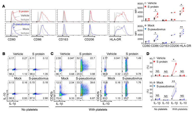
Figure 4. SARS-CoV-2–activated platelets induced monocyte differentiation toward a proinflammatory phenotype.
Purified monocytes from healthy donors were cocultured with purified autologous platelets in the presence of spike protein or S-pseudovirus, and then washed to remove nonadherent surplus platelets. (A) After another 48 hours in culture, the cell surface expression of CD80, CD86, CD163, CD206, and HLA-DR on monocytes was analyzed by flow cytometry (n = 3). (B–D) The expression of IL-1β and IL-10 in monocytes was examined by intracellular cytokine staining after 12 hours in culture, and CD42b indicated platelet-monocyte adherence in cocultures (n = 4). P value by paired Student’s t test is displayed. *P < 0.05; **P < 0.01.

