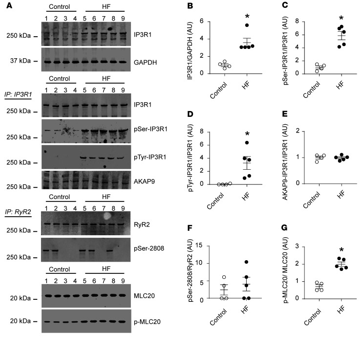
Figure 1. IP3R1 remodeling in aortic tissues from patients with HF.
(A) Representative immunoblots showing total IP3R1 expression, phosphorylation of serine/tyrosine kinases, and AKAP9 binding to the channels, as assessed by immunoprecipitation (IP: IP3R1); RyR2 phosphorylation levels of PKA-induced RyR2 phosphorylation on serine 2808 (pSer-2808); and p-MLC20 levels in aortic tissues from patients with HF (n = 5) and controls (n = 4). (B–G) Quantification of the immunoblots shown in A. Individual values with the mean ± SEM are shown. *P < 0.05 versus control, by 2-tailed Student’s t test.

