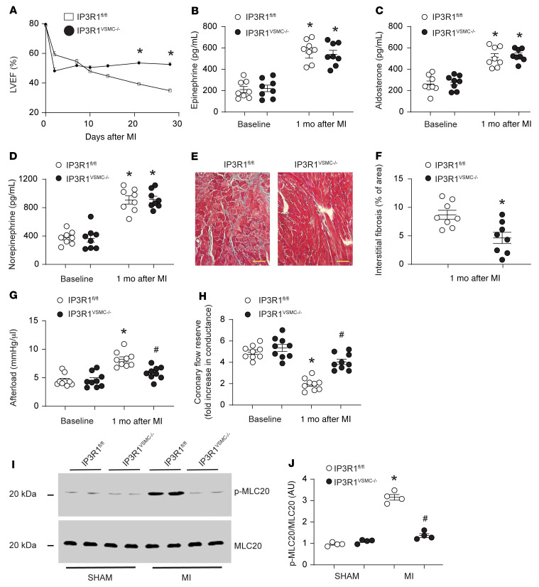
Figure 3. Functional role of VSMC IP3R1 in HF.
(A) LVEF evaluated by serial echocardiography following surgical ligation of the LAD. (B–D) Neurohormonal activation in HF assessed by measuring blood levels of catecholamines and aldosterone. (E) Representative Masson’s trichrome–stained images showing interstitial cardiac fibrosis. Scale bars: 50 μm. (F) Quantification of interstitial cardiac fibrosis. (G) Measurement of cardiac afterload (ratio of end-systolic pressure and stroke volume); other hemodynamic parameters are reported in Supplemental Table 2. (H) Coronary flow reserve determined in vivo in IP3R1fl/fl and IP3R1VSMC–/– mice. (I) Representative immunoblots of denuded mesenteric arteries (n ≥8 mice/group) showing MLC protein phosphorylation levels in sham-operated and MI IP3R1fl/fl and IP3R1VSMC–/– mice. (J) Quantification of results in I. Data are shown as individual values with the mean ± SEM. *P < 0.05 versus sham, by 2-tailed Student’s t test; #P < 0.05 versus MI IP3R1fl/fl mice, by repeated-measures ANOVA.

