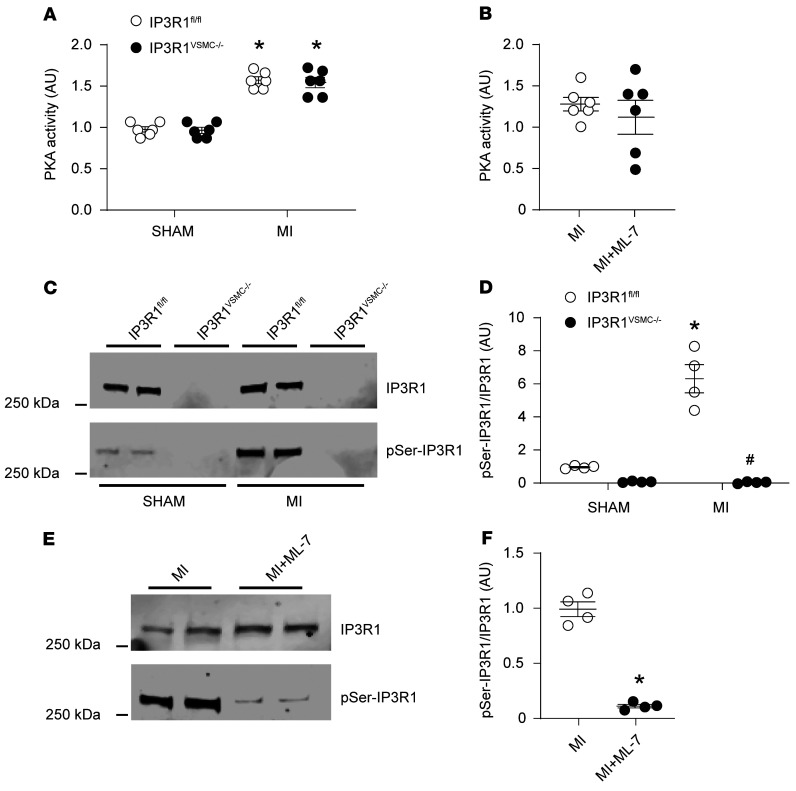
Figure 5. Increased IP3R1 phosphorylation by PKA in VSMCs during HF.
(A) PKA activity in VSMCs from sham-operated and MI IP3R1fl/fl and IP3R1VSMC–/– mice. (B) PKA activity in VSMCs from MI and ML-7–treated MI mice (n = 6 mice per group). (C and D) Representative immunoblots (C) and quantification (D) showing increased IP3R1 phosphorylation by PKA in VSMCs from IP3R1fl/fl mice after MI compared with sham-operated mice (n = 6 mice per group). (E and F) Representative immunoblots (E) and quantification (F) showing reduced IP3R1 phosphorylation by PKA in VSMCs from HF-treated mice compared with untreated mice (n = 4 mice per group). Individual values are shown with the mean ± SEM. *P < 0.05 for IP3R1fl/fl sham versus IP3R1fl/fl MI mice, IP3R1VSMC–/– sham versus IP3R1VSMC–/– MI mice, and MI mice versus ML-7–treated MI mice; 2-tailed Student’s t test. #P < 0.05, for IP3R1fl/fl MI versus IP3R1VSMC–/– MI mice; repeated-measures ANOVA.

