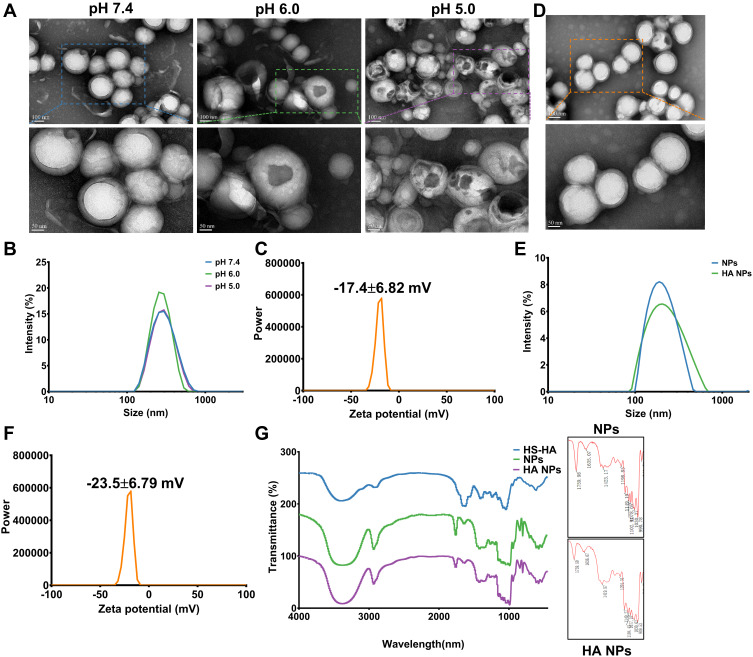
Figure 2.
Synthesis and characterization of “nano-bomb effect” nanoparticles and hyaluronic acid (HA) modified with nanoparticles. (A) TEM images of the nanoparticles under different pH conditions for 3 h at 37°C (n=5). The nanoparticles maintain a spherical morphology and core-shell structure at pH 7.4, and were broken open under lower pH (pH 6.0 and pH 5.0), indicated the pH-activated behavior. (B) Size distribution of nanoparticles at different pH conditions were measured by dynamic light scattering (DLS) (n=5). (C) The zeta potential of nanoparticles was −17.4±6.82 mV, which was measured by dynamic light scattering (DLS) (n=5). (D) TEM images of L-Arg-CO2@NPs modified with HA. (E and F) Size distribution and zeta potential of L-Arg-CO2@NPs (NPs) and HA-L-Arg-CO2@NPs (HA NPs) were determined by dynamic light scattering (DLS). (G) FT-IR spectra of HA-L-Arg-CO2@NPs (HA NPs) showing the changes of infrared spectra of the Michael addition reaction between maleimide and sulfhydryl.
Abbreviation: FT-IR, Fourier transform infrared spectroscopy.

