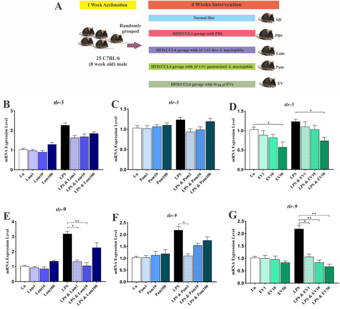
Figure 1.
(A) Study design of the animal experiment. Anti-inflammatory effects of all A. muciniphila supplementations in LX-2 cell line. The mRNA Level of tlr-5 in quiescence and LPS-activated LX-2 cells treated with (B) Lam (C) Pam and (D) EVs; and tlr-9 in quiescence and LPS-activated LX-2 cells treated with (E) Lam (F) Pam and (G) EVs. Un: untreated cells, Lam: live A. muciniphila, Pam: pasteurized A. muciniphila, EV: extra cellular vesicles of A. muciniphila. Data are expressed as mean ± SD (n = 5). *p < 0.05, **p < 0.01 and ***p < 0.001 by post hoc Turkey’s one-way ANOVA statistical analysis.

