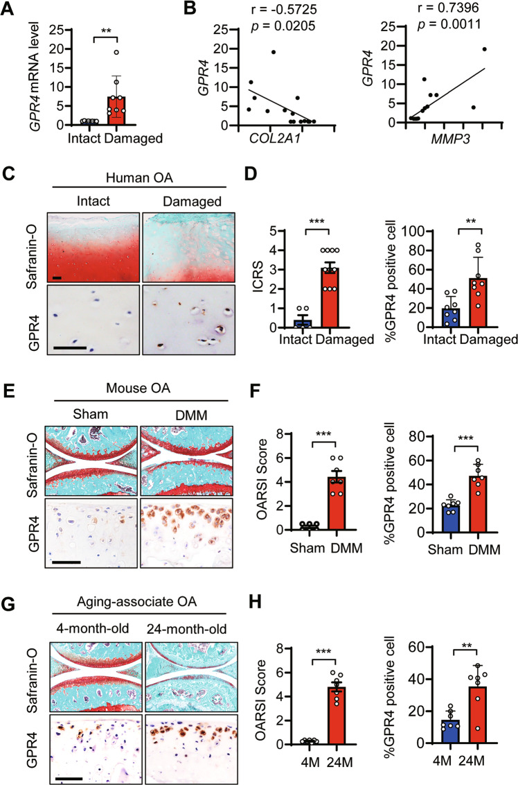
Fig. 1. GPR4 expression was upregulated in human and mouse OA articular cartilage.
A The mRNA levels of GPR4 in intact and damaged regions of articular cartilage from OA patients (n = 8) were determined by qRT-PCR. Data are expressed as mean ± s.d. **p < 0.01 by Student’s two-tailed t test. B Correlation between the expression of GPR4 and COL2A1 (left), or MMP3 (right) in intact and damaged regions of articular cartilage from OA patients (n = 8) by qRT-PCR. Pearson’s correlation analysis was performed. C, D Representative images of Safranin-O staining and immunohistochemistry (IHC) staining of GPR4 in intact and damaged regions of articular cartilage from human OA patients (C), Scale bar, 50 μm (n = 8 patients per group). International Cartilage Repair Society (ICRS) scores of human OA cartilage were analyzed (D, left) and GPR4 positive cells were quantified by IHC (D, right). n = 8. Data are expressed as mean ± s.d. **p < 0.01, ***p < 0.001 by Student’s two-tailed t test. E–H Staining of articular cartilage sections with Safranin-O and GPR4 IHC from sham-operated (n = 6) or DMM-operated (n = 7) mice (E) or 4- or 24-month-old mice (n = 7). Scale bars, 50 μm. G The corresponding OARSI (Osteoarthritis Research Society International) scores were assessed (F and H, left) and GPR4 expression was quantified (F and H, right), respectively. n = 7. Data are expressed as mean ± s.d. **p < 0.01, ***p < 0.001 by Student’s two-tailed t test.

