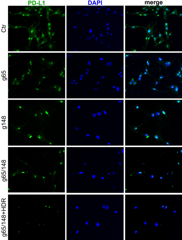
Figure 4.
Immunostaining of PD-L1. The cytoplasmic and nuclear PD-L1 were stained with antibody against PD-L1 (green). The nuclei was labeled with DAPI (blue). Dual immunofluorescence labeling of PD-L1 and DAPI in U87 cells treated to Cas9-g82, Cas9-g165, Cas9-g82/165, and Cas9-g82/165 + HDR.The untreated U87 cells served as control. Notable reduction of both cytoplasmic and nuclear PD-L1 was seen in U87 cells treated to Cas9-g82/165 + HDR.

