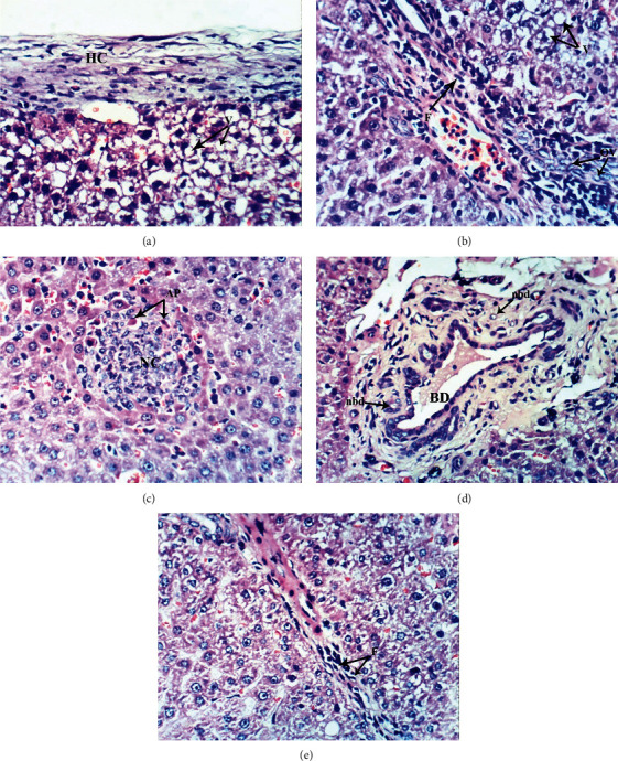
Figure 5.

Photomicrographs of liver sections of DOX-injected group showing thickening of the hepatic capsule (HC) and cytoplasmic vacuolization of hepatocytes (V) (a), fibroblast proliferation (F) in the portal tract and oval cell proliferation (OV) as well as vacuolization of hepatocytes (V) (b), focal hepatic necrosis (NC) as well as apoptosis of hepatocytes (AP) (c), hypertrophied bile duct (BD) and appearance of newly formed bile ductules (nbd) (d), and fibroblast proliferation (F) around the hepatocytes (e) (H & E ×400).
