
Figure 8.
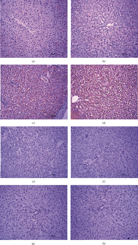
Photomicrographs (a–h) of immunhistochemical stained liver sections. (a, b) Normal rat liver showing weak immunohistochemical reaction of p53 expression. (c, d) Liver sections of DOX-injected rats showing a strong positive immunohistochemical reaction of p53 expression marked by dense brown color in cytoplasm and nucleus of hepatocytes. (e, f) Liver sections of DOX-injected rats treated with thyme oil showing a substantial decrease in the expression of p53. (g, h) Liver sections of DOX-injected rat treated with thymol showing a substantial decrease in the expression of p53.