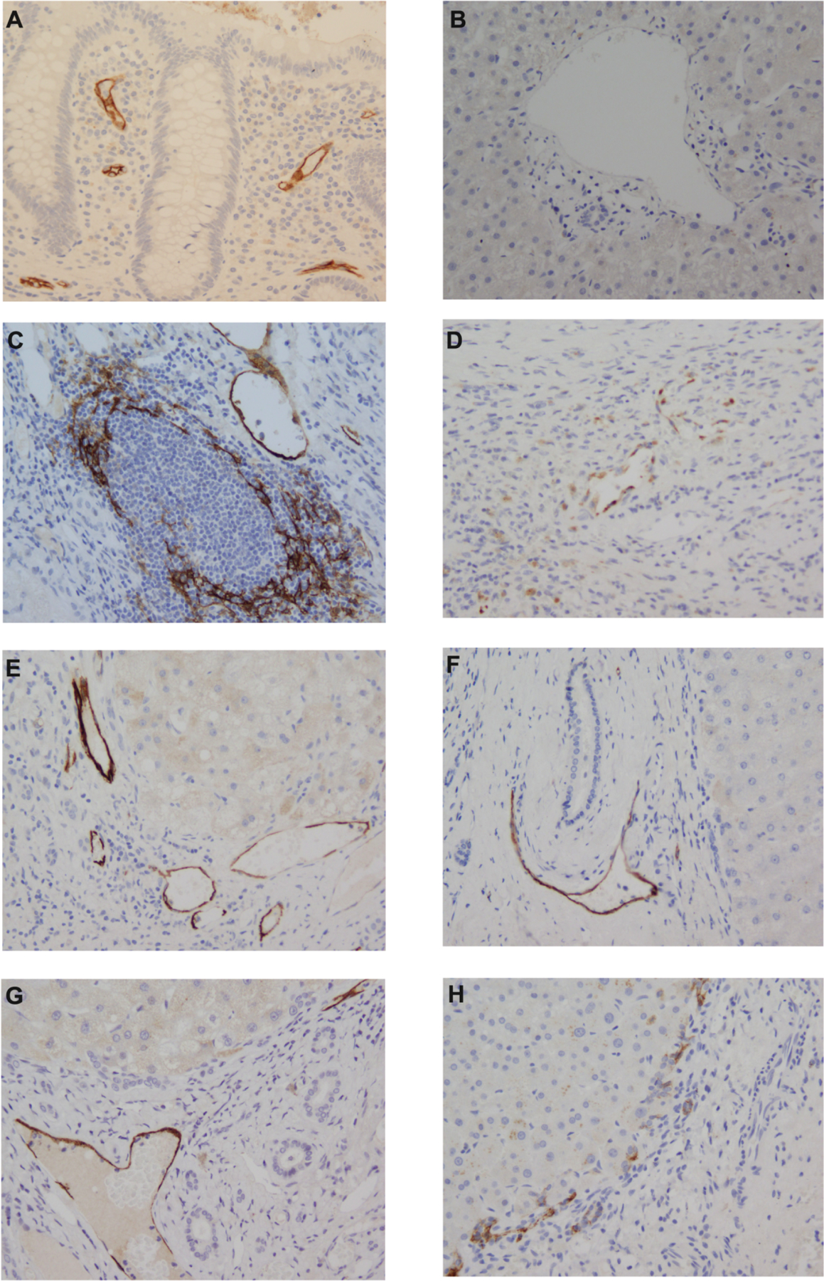
Figure 1. Representative MAdCAM-1 immunoreactivity patterns in patients with chronic liver disease explants.

[A] Colon control tissue; [B] Normal liver [C] chronic hepatitis C; [D] primary sclerosing cholangitis; [E] alcoholic liver disease; [F] primary biliary cholangitis; [G] non-alcoholic steatohepatitis; [H] Autoimmune hepatitis). Vessel MAdCAM-1 staining is present in panels C-G; lymphoid aggregate MAdCAM-1 staining in panel C (HCV patient. Similar patterns were also observed in PBC sections). No staining is present in panel A.
