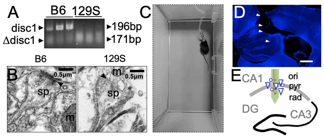
Figure 1: 129S mice exhibit spontaneous Disc1 mutation.

A, Gel electrophoresis demonstrating the amplified Disc1 gene in tail tissue biopsies of adult C57BL/6 and 129S mice. The mutant Disc1 gene in 129S mice shows truncation of the DNA sequence (179 bp) and decreased expression. The C57BL/6 strain expresses the wild-type Disc1 gene (196 bp).
B, Transmission electron photomicrograph showing decreased synaptic fidelity in the hippocampus in 129S mice, compared with a normal synaptic profile in the C57BL/6 hippocampus. Scale bar=0.5 μm. sp: dendritic spine, m: mitochondria, and black arrowhead: postsynaptic density).
C, A mouse with a neural electrode implant, head stage, and a tethered SPI cable.
D, Representative fluorescence image showing DAPI counterstaining of a brain slice obtained from a mouse with an electrode implant (scale bar=0.5 mm).
E, Illustration of neural probe placement in the CA1 of a mouse. Here, a linear array is shown for demonstration purposes. Other experiments included four channel microelectrode arrays (ori: oriens layer, pyr: pyramidal cell layer, and rad: radiatum layer of the CA1).
