
Figure 1.
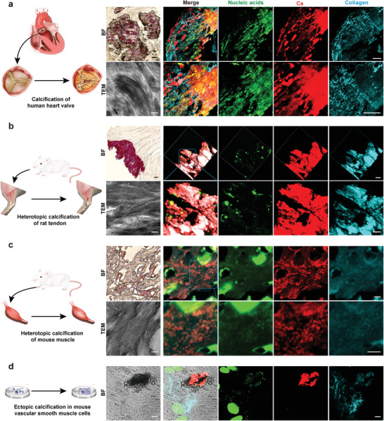
Extracellular DNA deposition identified from pathological collagen mineralization models in vivo and in vitro. a) Bright field (bar: 50 µm) and CLSM images (bar: 100 µm) taken from a human heart valve that was calcified in vivo showing the presence of randomly scattered extracellular DNA within the mineralized tissue. TEM image taken from the same specimen showing intrafibrillar mineralization of collagen fibrils within the tissues (bar: 200 nm). b) Bright field (bar: 50 µm) and CLSM images (bar: 20 µm) taken from an ectopically calcified rat Achilles tendon in vivo showing the presence of extracellular DNA within the mineralized tissue. TEM image taken from the same specimen showing intrafibrillar mineralization of collagen fibrils within the tissues (bar: 200 nm). c) Bright field (bar: 50 µm) and CLSM images (bar: 5 µm) taken from a bone‐like intramuscular ectopic calcification in vivo showing the presence of extracellular DNA within the mineralized tissue. TEM image taken from the same specimen showing intrafibrillar mineralization of collagen fibrils within the tissue (bar: 200 nm). d) Bright field (bar: 10 µm) and CLSM images (bar: 10 µm) taken from a mineralized nodule secreted by mouse vascular smooth muscle cells that were cultured in osteogenic medium in vitro. Similar to the three in vivo models, extracellular DNA was identified within the ectopically mineralized nodule.