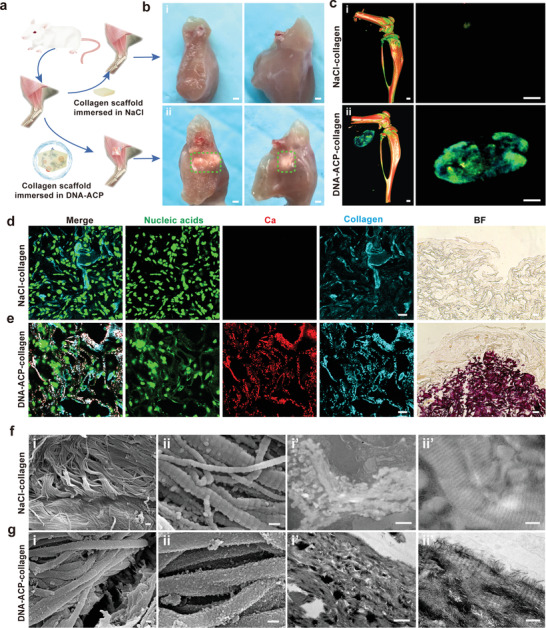
Figure 7.

DNA‐ACP induced intramuscular ectopic calcification in vivo. a) Scheme of the surgical operation in the in vivo murine intramuscular ectopic calcification experiment. b) Photograph of intramuscular ectopic calcification samples at 3 weeks post implantation (bars: 1 mm). c) 3D reconstruction of micro‐CT scans of the ectopic calcification samples (bars: 0.5 mm). b[i],c[i]) There was no sign of collagen mineralization in the NaCl control. b[ii],c[ii]) The DNA‐ACP group showed evident ectopic calcification in the muscle tissue. d) CLSM images of the NaCl control group showed no calcium deposition (bar: 20 µm). Bright field: optical image taken from the same specimen (bar: 50 µm). e) CLSM images of intramuscular ectopic calcification induced by DNA‐ACP collagen scaffolds (bar: 20 µm). Bright field: optical image of the same specimen (bar: 50 µm). f) SEM (f[i],[ii]) and TEM (f[i′],[ii′]) images of the sample from the NaCl control group showing unmineralized collagen fibrils (bars: i and i′: 1 µm; ii and ii′: 200 nm). g) SEM (g[i],[ii]) and TEM (g[i′],[ii′]) images of ectopic calcification induced by the DNA‐ACP‐dipped collagen scaffold. Both SEM and TEM images showed heavily extra/intrafibrillar collagen mineralization (bars: i and i′: 1 µm; ii and ii′: 200 nm).
