SUMMARY
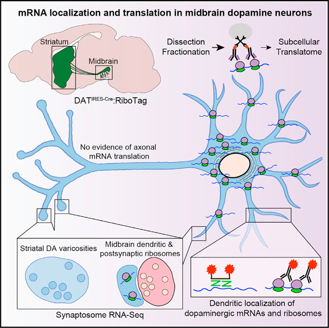
Midbrain dopaminergic (mDA) neurons exhibit extensive dendritic and axonal arborizations, but local protein synthesis is not characterized in these neurons. Here, we investigate messenger RNA (mRNA) localization and translation in mDA neuronal axons and dendrites, both of which release dopamine (DA). Using highly sensitive ribosome-bound RNA sequencing and imaging approaches, we find no evidence for mRNA translation in mDA axons. In contrast, mDA neuronal dendrites in the substantia nigra pars reticulata (SNr) contain ribosomes and mRNAs encoding the major components of DA synthesis, release, and reuptake machinery. Surprisingly, we also observe dendritic localization of mRNAs encoding synaptic vesicle-related proteins, including those involved in exocytic fusion. Our results are consistent with a role for local translation in the regulation of DA release from dendrites, but not from axons. Our translatome data define a molecular signature of sparse mDA neurons in the SNr, including the enrichment of Atp2a3/SERCA3, an atypical ER calcium pump.
In brief
Local translation regulates the subcellular proteome in neurons but has not been characterized in midbrain dopamine neurons, cells with large dendrites and axonal arborizations. Hobson et al. investigate messenger RNA localization and translation in midbrain dopamine neurons in the mouse brain, finding ribosomes and dopaminergic mRNAs in dendrites, but not axons.
Graphical Abstract
INTRODUCTION
Midbrain dopaminergic (mDA) neurons play critical roles in reward processing, movement control, and cognitive function. Their elaborate cytoarchitecture includes unmyelinated axons that course through the medial forebrain bundle (MFB) to reach basal ganglia and cortical targets (Björklund and Dunnett, 2007). Individual mDA neurons of the murine substantia nigra pars compacta (SNc) exhibit axonal arborizations reaching up to 500 μm in total length that possess 104–105 presynaptic varicosities (Matsuda et al., 2009). In addition to axonal DA release in the striatum and cortex, mDA neurons release DA within the midbrain (reviewed in Rice and Patel, 2015 and Cheramy et al., 1981), including from ventrally directed dendrites of SNc neurons that can project more than 500 μm into the substantia nigra pars reticulata (SNr) (Geffen et al., 1976; Korf et al., 1976; Tepper et al., 1987). Recent work has begun to identify molecular mechanisms that regulate DA release in the midbrain (Chen and Rice, 2001; Mendez et al., 2011; Robinson et al., 2019; Witkovsky et al., 2009) and striatum (Banerjee et al., 2020; Liu et al., 2018), but it is unclear how mDA neurons localize and maintain DA neurotransmission machinery in both dendritic and axonal compartments.
The subcellular proteome of neurons is regulated in part by local translation. Dendritic protein synthesis plays a critical role in several forms of postsynaptic plasticity (Bradshaw et al., 2003; Cracco et al., 2005; Huber et al., 2000; Kang and Schuman, 1996), and local translation is involved in the developmental pathfinding of axonal growth cones and the regeneration of peripheral axons (reviewed in Crispino et al., 2014; Jung et al., 2012). Recent evidence suggests that local translation occurs in mature central nervous system axons of excitatory and inhibitory neurons (Hafner et al., 2019; Ostroff et al., 2019; Scarnati et al., 2018; Shigeoka et al., 2016; Younts et al., 2016). Less is known about local translation in monoaminergic neurons. Intriguingly, the messenger RNA (mRNA) encoding tyrosine hydroxylase (TH), the rate-limiting enzyme in catecholamine biosynthesis, is localized to axons of sympathetic neurons in vitro (Gervasi et al., 2016). Ablation of an axonal localization motif in the 3′ untranslated region of Th mRNA decreases axonal TH protein levels and release of norepinephrine (Aschrafi et al., 2017), suggesting that local protein synthesis might regulate neurotransmission in mDA neurons.
Here, we systematically investigate mRNA localization and translation within mDA neurons in the mouse brain. Our results reveal the subcellular organization of protein synthesis in mDA neurons, with implications for the regulation of DA neurotransmission in health and disease.
RESULTS
DATIRES−Cre:RiboTag mice enable visualization and capture of mDA neuronal ribosomes
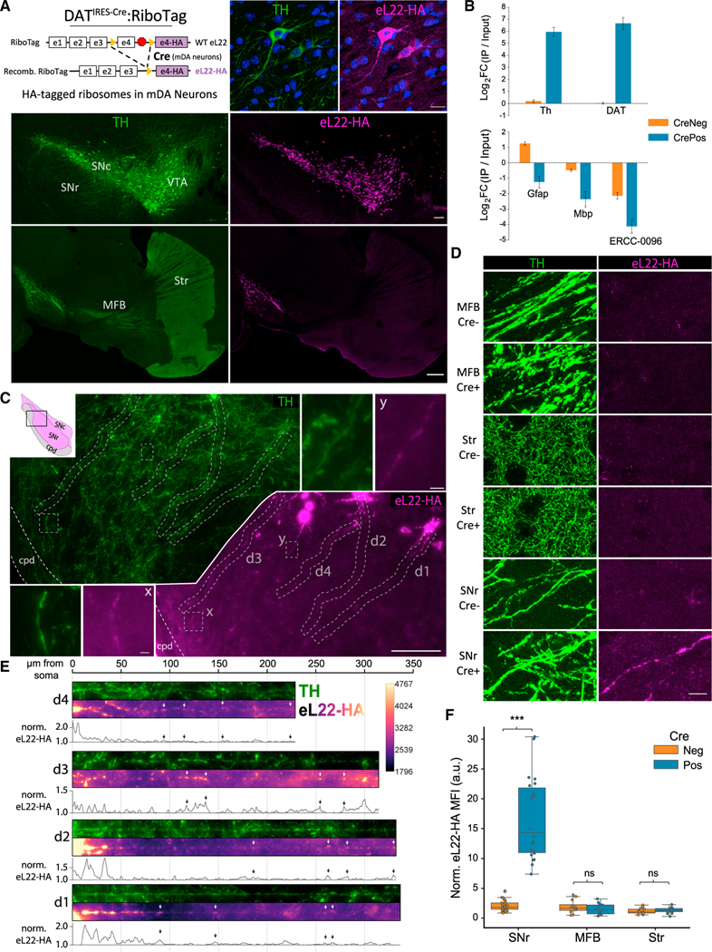
To study subcellular translation in mDA neurons, we crossed DATIRES−Cre mice (Bäckman et al., 2006) with RiboTag mice (Sanz et al., 2009) to express HA-tagged eukaryotic ribosomal protein L22 (eL22-HA) specifically in TH+ mDA neurons in the SNc and ventral tegmental area (VTA) (Figures 1A and S1A). Anti-HA immunoprecipitation (IP) of ventral midbrain (VM) polysome extracts from Cre+ RiboTag mice (DATIRES−Cre/wt; RiboTag+/−) yielded a more than 64-fold enrichment of mDA neuron-specific mRNAs Th and Slc6a3/DA transporter (DAT), and depleted glial and mRNAs Gfap, Mbp, and the soluble poly-adenylated spike-in standard ERCC-0096 (Figure 1B).
Figure 1. Subcellular distribution of eL22-HA tagged ribosomes in adult (10–14 mo) DATIRES−Cre:RiboTag mice.
(A) DATIRES−Cre:RiboTag genetics (upper left). TH and eL22-HA IF. Upper right: mDA neurons in the SNc, DAPI in blue. Scale bar, 20 μm. Middle: Coronal midbrain section. Scale bar, 100 μm. Lower: Sagittal section. Scale bar, 500 μm.
(B) qRT-PCR from VM Input and RiboTag IPs from DATIRES−Cre:RiboTag mice (Cre+, n = 7) or Cre− littermates (n = 5) showing IP/Input enrichment relative to Actb (mean ΔΔCq +/− SEM).
(C) Ventrolateral SNc and SNr stained for TH and eL22-HA. x, y insets shown in white lines. Dendrites d1-d4 are displayed below in (E). Scale bars, main image, 100 μm; insets, 5 μm. cpd, cerebral peduncle.
(D) TH and eL22-HA staining in the MFB, striatum, and SNr of Cre−/Cre+ RiboTag mice. Scale bar, 10 μm.
(E) Straightened dendritic segments d1-d4 from (C) with eL22-HA intensity normalized to local background and plotted below. Arrows indicate eL22-HA hotspots.
(F) Quantification of eL22-HA signal within TH+ neurites in the MFB (axons), striatum (axons), and SNr (dendrites) of Cre− and Cre+ mice. Data are background-normalized mean eL22-HA intensity of TH+ pixels within a field of the indicated region (n = 6–10 fields, n = 4 sections, n = 3 mice per each genotype/region). Two-way ANOVA main effects: Region, F = 74.4 (2, 100), p = 1.6−20; genotype, F = 117.2 (1, 100), p = 1.5 3 10−18; and region:fenotype interaction, F = 68.4 (2,100), p = 1.9 3 10−19. Tukey’s HSD post hoc test for Cre+ versus Cre− staining: SNr (p-adj < 0.001), MFB and Str (both p-adj > 0.9). ***p-adj < 0.001.
Consistent with previous studies of DAT-Cre lines (Bäckman et al., 2006; Lammel et al., 2015; Mingote et al., 2017; Turiault et al., 2007), no expression of eL22-HA was observed in the dorsal striatum, nucleus accumbens, or cortex (Figure 1C). In concordance with recent work using another DAT-Cre line (Papathanou et al., 2019), we found scattered TH−/tdTom+ cells in the lateral septum of DATIRES−Cre/wt mice crossed to Ai9 tdTomato reporter mice (Figure S1B) (Madisen et al., 2010). However, we found no evidence of eL22-HA expression in the bed nucleus of stria terminalis or lateral septum of DATIRES−Cre/wt;RiboTag+/− mice (Figure S1B). Nonetheless, we removed all tissue medial to the lateral ventricles (see STAR Methods) in our striatal dissections of DATIRES−Cre/wt;RiboTag+/− mice to ensure that striatal eL22-HA was derived from solely from mDA axons.
We analyzed the subcellular distribution of mDA neuronal ribosomes using anti-HA immunofluorescence (IF). eL22-HA labeling was present in soma and proximal dendrites (Figures 1A and S1C), but undetectable within axons in the MFB or striatum (Figure 1A). SNc mDA neurons typically possess three to six long, mostly unbranched dendrites; the largest one or two are directed ventrolaterally into the SNr (Juraska et al., 1977; Prensa and Parent, 2001; Tepper et al., 1987). With tyramide signal amplification (Adams, 1992; Bobrow et al., 1992) (Figures S1D and S1E), eL22-HA labeling was apparent within such dendrites in the SNr (Figures 1C and 1E), even at the distal edge near the cerebral peduncles (cpd; Figure S1F, lower). Co-localization with TH IF confirmed that eL22-HA clusters were scattered throughout ventral-directed dendrites of SNc mDA neurons (Figures 1C and 1E), which could be distinguished from a few mDA neuronal soma in the SNr (Figure S1F, upper). Consistent with a previous study (Brichta et al., 2015), we observed no specific eL22-HA labeling in the MFB or striatum (Figure 1D). Quantification of eL22-HA in Cre+ versus Cre− (DATwt/wt;RiboTag+/−) mice revealed significantly higher fluorescence in TH+ SNr dendrites, but not TH+ axons in the MFB or striatum (Figures 1D and 1F). Assuming that eL22-HA reflects the localization of endogenous ribosomes (Sanz et al., 2009; Shigeoka et al., 2016), these data are consistent with the majority of mDA neuronal ribosomes residing in the soma and proximal dendrites, with lesser abundance in the distal dendrites and exceedingly low levels in axons.
Sensitive, quantitative capture of dopaminergic ribosomes from regional dissections
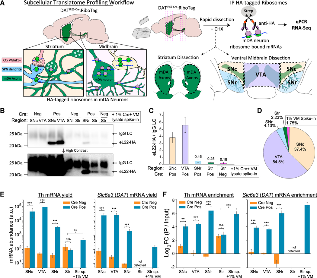
To identify translating mRNAs in distinct subcellular compartments of mDA neurons, we conducted RiboTag IP on dissections of four regions (Figure 2A): (1) the dorsal and ventral striatum, containing mDA axons, (2) the VTA, containing mDA somata and dendrites, (3) the SNc, containing mDA somata and dendrites, and (4) the SNr, containing a few mDA somata amid a high density of ventral-projecting dendrites of SNc mDA neurons (Figure S1G). Owing to the low yields from axonal RiboTag, we used Cre− mice to control for non-specific binding (Shigeoka et al., 2016). The total RNA yield from Cre− IPs was typically 10s–100s of picograms as estimated using qualitative reverse transcriptase polymerase chain reaction (qRT-PCR) for b-actin (Actb) (Figure S1I).
Figure 2. Regional distribution of eL22-HA protein and dopaminergic mRNAs captured by RiboTag IP in adult mice (10–14 mo).
(A) Subcellular translatome profiling workflow.
(B) Western blot of captured eL22-HA from RiboTag IPs. eL22-HA (23 kDa) is detected below IgG light chain (LC, approximately 25 kDa). Lower: High contrast reveals bands in striatal samples and 1% VM spike-in.
(C) Quantification of Western blot eL22-HA, normalized to IgG LC with mean ± SEM shown for each region/genotype (n = 3–4).
(D) Fractional abundance of eL22-HA captured in each region, using normalized eL22-HA intensity from (C).
(E and F) qRT-PCR of Th and Slc6a3/DAT mRNA in RiboTag IPs from each region/genotype (n = 3–4 each). (E) mRNA abundance in arbitrary units (240–Cq). Mean (A)u. +/− SEM are plotted. For Th mRNA, two-way ANOVA main effects: region, F = 17.7 (3, 19), p = 9.8 3 10−6; genotype, F = 168.1 (1, 19), p = 6.9 3 10−11; and region:genotype interaction, F = 20.1 (3, 19), p = 4.0 3 10−6. Tukey’s HSD post hoc for Cre+ versus Cre− samples: ***p-adj<0.001 for SNc, VTA, and SNr, but not for Striatum (p-adj > 0.9). Welch’s t test for 1%VM/Str_Sp samples versus Cre+/Cre− striatal samples: t(9) = 3.72, p = 0.0062. For DAT mRNA, two-way ANOVA main effects: region, F = 13.0 (2, 15), p = 5.3 3 10−4; genotype, F = 262.5 (1, 15), p = 6.5 3 10−11; and region:genotype interaction, F = 1.46 (2, 15), p = 0.26. DAT mRNA was not detected in striatal samples. Tukey’s HSD post hoc test for Cre+ versus Cre− samples: p-adj < 0.001 for the SNc, VTA, and SNr (***). (F) qRT-PCR for RiboTag IP/Input enrichment relative to Actb (mean ΔΔCq +/− SEM). For Th mRNA, two-way ANOVA main effects: genotype, F = 154.5 (1, 20), p = 7.3 3 10−11; region, F = 1.97 (3, 20), p = 0.15; and region:genotype interaction, F = 23.4 (3, 20), p = 9.4 3 10−7. Tukey’s HSD post hoc test for Cre+ versus Cre− samples: p-adj < 0.01 for SNc, VTA, and SNr, but not for striatum (p-adj > 0.9). Welch’s t test for 1%VM/Str_Sp samples versus Cre+/Cre− striatal samples: t(10) = 6.66, p = 0.0009. For Slc6a3/DAT mRNA, two-way ANOVA main effects: genotype, F = 269.2 (1, 15), p = 5.4 3 10−11; region, F = 1.96 (3, 15), p = 0.18; and region:genotype interaction, F = 19.1 (2, 15), p = 7.5 3 10−5. Tukey’s HSD post hoc test for Cre+ versus Cre− samples: p-adj < 0.001 for SNc, VTA, and SNr (**p-adj < 0.01, ***p-adj < 0.001).
We estimated eL22-HA abundance in each dissected region by Western blotting eL22-HA IPs. To estimate the sensitivity of our striatal IP, we included control samples of Cre− striatal lysates spiked with 1% of VM lysates from Cre+ mice. eL22-HA bands were prominent in VTA and SNc IPs, while faint eL22-HA signal was only visible at high contrast in IPs from the striatum and 1% VM spike-in control (Figures 2B and 2C). Quantification revealed that of all eL22-HA captured, 37.4% was from the SNc, 54.5% from the VTA, 4.13% from the SNr, 2.23% from the striatum, and 1.75% from our 1% VM spike-in control (Figure 2D). Striatal eL22-HA abundance was not significantly different from the 1% VM spike-in control. eL22-HA abundance correlated well with our spike-in control (1% of VM lysate vs. an estimated 1.75% eL22-HA) and the reported distribution of mDA neurons in C57BL/6J mice (Nelson et al., 1996): approximately 8,000 in the SNc (38% of mDA neurons vs. 37.4% of eL22-HA) and approximately 10,000 in the VTA (47.6% of mDA neurons vs. estimated 54.5% of eL22-HA). The eL22-HA Western blot and histology data support the soma as the major site of protein synthesis in mDA neurons (Palay and Palade, 1955) and are consistent with very low levels or the absence of ribosomes in striatal mDA axons.
qRT-PCR of RiboTag IPs revealed significant Cre-dependent increases in yield and enrichment of Th and Slc6a3/DAT mRNA in IPs from SNc and VTA (Figures 2E and 2F). We also found a significant Cre-dependent increase in the yield of Th and Slc6a3/DAT mRNAs in SNr IPs (Figure 2E) and an enrichment of Th and Slc6a3/DAT from the SNr that was higher than in the SNc and VTA (Figure 2F). When comparing striatal IPs from Cre+ and Cre− mice, we found no significant differences in Th mRNA yield or enrichment, and Slc6a3/DAT was undetectable in all samples (Figures 2E and 2F). Similar to the SNr, we found a more than 64-fold Cre-dependent enrichment of both Th and Slc6a3/DAT in our 1% VM spike-in control, despite a low yield (Figures 2E and 2F). The yield and enrichment of Th mRNA were significantly higher in our 1% VM spike-in controls than striatal IPs from Cre− and Cre+ mice (Figures 2E and 2F). These results demonstrate sensitive, quantitative RiboTag IP from distinct mDA neuron compartments and suggest that Th and Slc6a3/DAT mRNAs are translated in dopaminergic dendrites in the SNr, but not in striatal mDA axons (Table 1).
Table 1.
Summary of RiboTag IP capture for eL22-HA, Th mRNA, and Slc6a3/DAT mRNA
| Percentage of total yield | ||||
|---|---|---|---|---|
|
| ||||
| Region | eL22-HA protein | Th mRNA |
Slc6a3/DAT mRNA |
|
| SNc | 37.4 ± 8.2% | 25.5 ± 6.4% | 42.0 ± 5.2% | |
| VTA | 54.5 ± 8.8% | 69.6 ± 5.4% | 52.9 ± 4.1% | |
| SNr | 4.13 ± 0.77% | 4.0 ± 1.2% | 4.62 ± 1.02% | |
| Striatum | 2.23 ± 0.33% | 0.14 ± 0.07% | Undetectable | |
| Striatum +1% VM spike-in | 1.75 ± 1.24% | 0.78 ± 0.52% | 0.51 ± 0.29% | |
| TH (Th) mRNA | ||||
|
| ||||
| Region | Log2 (ΔYield) Cre+ - Cre− | Tukey HSD (p-adj) | Log2 FC (IP/Input) Cre+ - Cre− | Tukey HSD (p-adj) |
|
| ||||
| SNc | 7.88 ± 1.16 | <0.001 | 2.88 ± 0.44 | 0.0013 |
| VTA | 10.49 ± 0.73 | <0.001 | 4.28 ± 0.52 | <0.001 |
| SNr | 6.93 ± 1.03 | <0.001 | 6.82 ± 0.73 | <0.001 |
| Striatum | −1.16 ± 1.25 | >0.9 | 0.20 ± 0.50 | >0.9 |
| Striatum +1% VM spike-in | 1.94 ± 0.94 | NA | 5.89 ± 0.40 | NA |
| Dopamine transporter (Slc6a3/DAT) mRNA | ||||
|
| ||||
| Region | Log2 (ΔYield) Cre+/Cre− | Tukey HSD (p-adj) | Log2 FC (IP/Input) Cre+ - Cre− | Tukey HSD (p-adj) |
|
| ||||
| SNc | 8.41 ± 1.30 | <0.001 | 3.62 ± 0.51 | <0.001 |
| VTA | 9.65 ± 0.78 | <0.001 | 3.54 ± 0.22 | <0.001 |
| SNr | 7.45 ± 0.48 | <0.001 | 7.48 ± 0.78 | <0.001 |
| Striatum | NA | NA | NA | NA |
| Striatum +1% VM spike-in | Cre− undetectable | NA | 7.21 ± 0.44 (Cre+) Cre− undetectable | NA |
Upper: Mean percentage of total yield ±SEM for each region across all Cre+ RiboTag IPs. Data from Figures 2B–2D (eL22-HA protein, n = 3 each region) and Figures 2E and 2F (Th and Slc6a3 mRNA, n = 3–4 each genotype/region).
Lower: Mean log2 differences in yield (left) or enrichment (right) ± SEM between Cre+ and Cre− RiboTag IPs for each region/mRNA. Data and p-adj from Tukey’s HSD post hoc comparisons are from Figures 2E and 2F (Th and Slc6a3 mRNA, n = 3–4 each genotype/region).
Lack of evidence for axonal translation in striatal RiboTag IPs
To identify mRNAs bound to putative axonal ribosomes, we analyzed the content of striatal RiboTag IPs from Cre− and Cre+ samples using RNA sequencing (RNA-seq). To accommodate picogram samples, we used a low input, pooled, 3′-end library construction strategy designed for single-cell RNA-seq (scRNA-seq) (Snyder et al., 2019) that includes unique molecular identifiers (UMIs) to mitigate PCR bias and estimate the number of transcripts captured per gene. Developing axons typically have a greater translational capacity than mature axons, which may reflect the downregulation of axonal ribosomes after synaptogenesis (Costa et al., 2019). Thus, in addition to middle-aged adult mice (10–14 months of age) (Figure 2), we also conducted RiboTag IPs from the striatum of Cre+ and Cre− mice at postnatal ages P0, P7, P14, P21, P31, and P90 (69 mice total, n = 2–7 each for Cre− and Cre+ mice at each age). We used a generalized linear model (GLM) within DESeq2 (Love et al., 2014) to test whether mRNAs were significantly enriched in IP versus Input samples only in Cre+ mice (Figure S2A) (see STAR Methods).
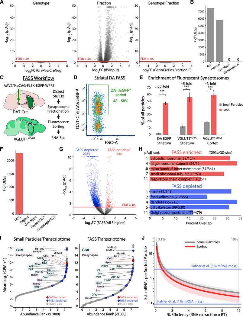
We found no significant effect of genotype for any genes (Figures 3A and 3B), indicating that the single DATIRES−Cre allele does not alter the striatal transcriptome. However, there was a significant effect of fraction for more than 5,000 genes (Figures 3A and 3B), demonstrating conservation of IP versus the input sample differences across genotype and age. This reflects age- and genotype-independent bias in non-specific binding of striatal polysome lysates during RiboTag IP. We found a significant effect of age for more than 7,000 genes (Figure 3B), consistent with developmental regulation of the striatal transcriptome that is conserved across fraction and genotype. Critically, we found no significant genotype:fraction interaction for any gene (Figures 3A and 3B), and thus found no statistical evidence for age-conserved, Cre-dependent axonal RiboTag IP enrichment of any genes.
Figure 3. Striatal RiboTag IP and dopaminergic synaptosome sorting provide no evidence of translation in DA axons.
Data are from P0 to adult mice in (A), (B) and adult (10–14 mo) for (C–J).
(A) DESeq2 GLM analysis of striatal RiboTag IP and Input samples from Cre-/Cre + mice: (P0: n = 6/6, P7: n = 6/6, P14: n = 6/6, P21: n = 6/7, P31: n = 2/2, P90: n = 2/3, 10–14 mo: n = 6/4, with IP and Input samples for each mouse. Left: genotype effect across levels of fraction (IP and Input). Middle: fraction effect across levels of genotype (Cre+ and Cre−). Right: genotype:fraction interaction. log2(GenoCrePos.FractionIP) is the difference in fraction effect between genotypes: {Cre+ log2FC(IP/Input) – Cre− log2FC(IP/Input)}. See Figure S2A schematic.
(B) Number of DEGs (FDR < 0.05) from DESeq2 in (A).
(C) FASS RNA-seq schematic. (D) Density plot of EGFP-sorted striatal synaptosomes from DAT-Cre mice expressing EGFP in mDA neurons.
(E) Comparison of fluorescent particles in unsorted and sorted synaptosome samples. Mean ± SEM for the %EGFP+/VENUS+ out of all particles are plotted for: DAT:EGFP striatum (n = 6), VGlut1VENUS striatum (n = 3), and VGlut1VENUS cortex (n = 3). Two-way ANOVA main effects: region/genotype, F = 16.9 (2, 18), p = 7.3 × 10⁻⁵; fraction, F = 427.9 (1, 18), p = 5.4 3 10−14; and region/genotype:fraction interaction, F = 0.30 (2, 18), p = 0.74. Tukey’s HSD post hoc test for unsorted versus sorted samples: p-adj < 0.001 for all three comparisons. ***p < 0.001.
(F) Number of DEGs (FDR < 0.05) from the DESeq2 with indicated terms removed from the GLM: ~FASS + region + genotype + region:FASS + genotype:FASS.
(G) Volcano plot for DESeq2, comparing FASS samples to all small particles (enriched/depleted genes, FDR < 0.05).
(H) GO analysis of FASS-enriched and -depleted DEGs from (G).
(I) Abundance versus rank for all small particles (left) and FASS samples (right). FASS-enriched and -depleted mRNAs are shown in red and blue, respectively.
(J) Estimated mRNAs per sorted particle as a function of FASS RNA-seq efficiency with estimates for whole forebrain VGlut1VENUS sorted particles from Hafner et al., 2019 (see STAR Methods).
We conducted the same analyses at each age, again finding no significant effect of genotype or genotype:fraction interaction, but a significant effect of fraction for more than 1,000 genes at most ages (Figure S2B). These findings are consistent with undetectable Cre-dependent mRNA capture in all striatal RiboTag IPs, regardless of age. Furthermore, we found no significant Cre-dependent differences in Th or Actb yield (Figure S2C) or in total mRNA yield as determined by total UMIs per sample (Figure S2D). Thus, despite our RiboTag IP and RNA-seq protocol with single-cell sensitivity, we found no evidence of ribosome-bound mRNAs derived from DA axons in the striatum. The yield from Cre− striatal IPs was on the order of 1–50 cells (10–500 pg total RNA via qRT-PCR and 20,000–200,000 UMIs via RNA-seq), and thus the yield of axonally derived ribosomes likely falls below this background level. We conclude that translating ribosomes in striatal DA axons are extremely sparse and not amenable to detection using striatal RiboTag IP.
Lack of dopaminergic mRNA signature in sorted striatal synaptosomes
Another approach to study the presynaptic transcriptome involves enrichment of resealed nerve terminals containing a genetically encoded fluorescent reporter (Biesemann et al., 2014; Hafner et al., 2019; Luquet et al., 2017). To directly interrogate the transcriptome of dopaminergic synaptosomes, we used our recently developed DA fluorescence activated synaptosome sorting (FASS) protocol (Paget-Blanc et al., 2021), where mDA neurons are labeled by injection of adeno-associated virus (AAV) expressing Cre-dependent eGFP into DAT-Cre mice (Figure 3C). We gated on small particles to avoid synaptosomal aggregates (Biesemann et al., 2014; Hobson and Sims, 2019) and sorted DAT:EGFP+ particles (Figure S2E). Reanalysis revealed 43%–58% EGFP+ particles in the sorted samples (Figure 3D), reflecting a more than 20-fold enrichment of DAT:EGFP+ synaptosomes (Figures 3E and S2E). However, resealed axonal varicosities of mDA neurons can remain stably bound to VGlut1+ presynaptic boutons and other synaptic elements (Paget-Blanc et al., 2021). To control for mRNA derived from co-enrichment of these other synaptic elements, we also sorted VGlut1+ synaptosomes from the striatum and cortex of VGlut1VENUS mice (Figure 3C). VGlut1VENUS+ particles were more abundant than DAT:EGFP+ particles in striatal synaptosomes, resulting in a slightly higher sorted purity (50%–60%), but substantially lower enrichment (approximately five-fold) of VGlut1VENUS+ synaptosomes compared with DAT:EGFP+ synaptosomes (Figures 3E and S2E). We also sorted all small particles from each sample to control for bias owing to the small particle gating and sorting procedure.
We used the same RNA-seq protocol as above to characterize the transcriptome of FASS and small particle sorted samples. For each sample of 1.5–7.5 million particles, we obtained between 104–105 UMIs (Figure S2F). For striatal samples, DAT:EGFP and VGlut1VENUS FASS samples yielded significantly fewer UMIs per sorted particle compared with all small particles (Figure S2G), although the yield from both sample types was only approximately 1 UMI per 50–200 sorted particles. A principal component analysis (PCA) clearly separated FASS samples from small particle controls along PC1, but DAT:EGFP FASS samples were not separated from striatal or cortical VGlut1VENUS FASS samples (Figures S2H and S2I). As above, we used DESeq2 to identify differentially expressed genes (DEGs) across FASS and control samples (see STAR Methods). We found a significant effect of FASS for more than 2,000 genes, but no significant effect of region:FASS or genotype:FASS interactions for any gene (Figure 3F). These data demonstrate that the FASS transcriptome is distinct from all small particles, but that the VGlut1VENUS and DAT:EGFP sorted samples are largely indistinguishable. Given the paucity of DA axons in the cortex compared to the striatum, this result argues against any detectable contribution of DA synaptosome-specific mRNAs. Most of the 240 genes enriched in FASS samples were enriched by more than 2-fold (Figure 3G), substantially lower than the more than 20-fold enrichment of DAT-EGFP+ particles in FASS sorted samples. No canonical dopaminergic mRNAs (e.g., Th, Slc6a3/ DAT) or other DA neuron-specific mRNAs were detected in any FASS samples.
Presynaptic elements are often bound to other particles in synaptosome preparations, which can represent non-specific aggregation (Hobson and Sims, 2019) or native adherence of synaptic structures (Paget-Blanc et al., 2021). Astrocytic processes containing mRNA and ribosomes are also present in synaptosome preparations (Chicurel et al., 1993; Mazaré et al., 2020; Sakers et al., 2017) and are a likely source of mRNA in our FASS samples. We found that, despite a relative depletion by sorting, many oligodendrocyte- and astrocyte-enriched mRNAs were abundant in our striatal FASS samples, while the microglia- and astrocyte-enriched mRNAs Cst3 and Apoe were enriched by sorting (Figure 3I). Many mDA axonal varicosities lack active zone scaffolding proteins and do not release DA upon stimulation (Liu et al., 2018; Pereira et al., 2016). We found no evidence for the local translation of active zone proteins in dopaminergic synaptosomes: Rims1 and Bsn mRNAs had low abundance in small particle samples and were further depleted by sorting (Figure 3I), suggesting that local translation does not regulate active and silent presynaptic sites in mDA axons.
A gene ontology (GO) analysis revealed that mRNAs encoding axonal, dendritic, and cytoskeletal proteins were overrepresented among the FASS-depleted mRNAs, while mRNAs encoding ribosomal proteins and nuclear-encoded mitochondrial proteins were overrepresented among FASS-enriched mRNAs (Figure 3H). Although the latter two groups of mRNAs have been observed in axons (Aschrafi et al., 2016; Briese et al., 2016; Gumy et al., 2011; Shigeoka et al., 2016, 2019; Taylor et al., 2009), they are also present in dendrites (Fusco et al., 2021; Perez et al., 2021), the synaptic neuropil (Biever et al., 2020; Cajigas et al., 2012) and perisynaptic astrocyte processes (Mazaré et al., 2020). Thus, it is unclear whether any FASS-enriched mRNAs are derived from DA synaptosomes. Since millions of synaptosomal particles yielded UMI counts similar to a single cell (Figure S2F), we estimated the mRNA content of sorted synaptosomal particles (see STAR Methods). Based on estimates of RNA extraction and reverse transcription efficiency, we estimate there are between 0.2 and 2 mRNAs per sorted synaptosomal particle (Figure 3J), consistent with the total RNA yield from VGlut1VENUS FASS samples (Hafner et al., 2019) of 0.1–2.2 mRNAs per sorted synaptosomal particle (Figure 3J). In addition to the lack of a DAT:EGFP-specific signature and a major contribution of glial mRNAs to the striatal FASS transcriptome, it is possible that many striatal synaptosomes contain no mRNA. Collectively, these data provide no evidence for mRNA localization in dopaminergic synaptosomes.
In a final effort to enrich mRNAs from mDA axons, we conducted RiboTag IP on striatal synaptosomes from DATIRES−Cre: RiboTag mice. Similar to whole striatal IPs, we observed no significant effect for genotype or genotype:fraction interaction, while hundreds of genes were significantly affected by fraction (Figure S2J). We found no significant difference in the mRNA yield of Cre+ versus Cre− IPs (Figure S2K), and there was no Cre-dependent bias for FASS-enriched or -depleted mRNAs (Figures S2L and S2M). Collectively, these data strongly suggest that mRNAs enriched in striatal FASS samples are not derived from ribosomes in dopaminergic varicosities.
mRNAs encoding DA transmission machinery are robustly localized to dopaminergic dendrites, but not axons
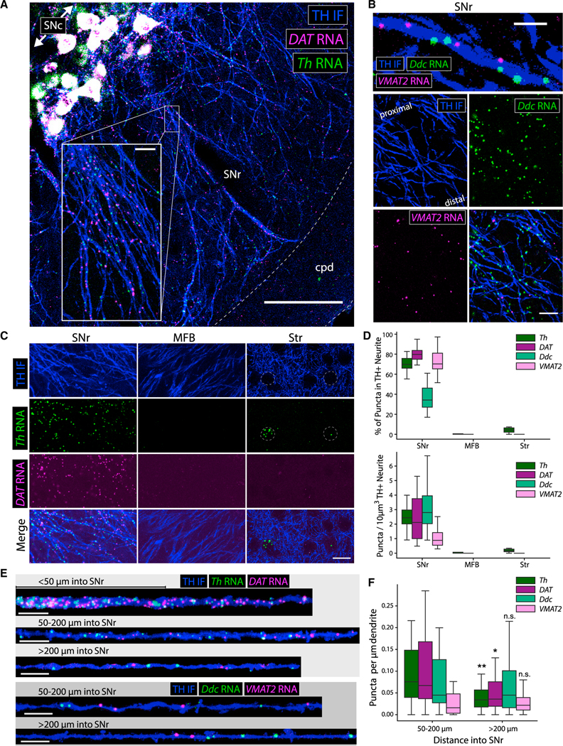
The eL22-HA staining in SNr dendrites (Figure 1) and enrichment of Th and Slc6a3/DAT mRNA in SNr RiboTag IPs (Figure 2) suggest local translation of these mRNAs in dopaminergic dendrites. We confirmed the dendritic localization of Th and Slc6a3/DAT mRNA using fluorescence in situ hybridization (FISH) (RNA-scope). In addition to dense staining of somata in the SNc, we observed dispersed Th and Slc6a3/DAT mRNA puncta throughout the SNr (Figure S3A). No staining was observed for DapB, a negative control bacterial mRNA (Figure S3B). Combining FISH with TH IF revealed a striking density of Th and Slc6a3/DAT mRNA puncta within TH+ dendrites in the SNr (Figure 4A). We found that Slc18a2/VMAT2 (vesicular monoamine transporter 2) and Ddc (aromatic l-amino acid decarboxylase) mRNAs were also localized in dopaminergic SNr dendrites (Figure 4B). Among these four dopaminergic mRNAs, only Ddc is expressed by other midbrain cells (Figure S3C), but Ddc puncta within TH+ dendrites were clearly distinguished from SNr cells (Figures S3D and S3E). All four mRNAs were observed within dopaminergic dendrites deep in the SNr, hundreds of microns from SNc soma (Figures 4A and S3F).
Figure 4. FISH reveals dendritic, but not axonal, localization of dopaminergic mRNAs in adult (10–14 mo) mouse brain.
(A) TH IF and FISH for Th and Slc6a3/DAT mRNA in the SN, cpd: cerebral peduncle. Scale bar, 100 μm; inset scale bar, 10 μm.
(B) TH IF and FISH for Ddc and Slc18a2/VMAT2 mRNA in the SNr. Scale bars, 5 μm (upper), 15 μm (lower).
(C) TH IF and FISH for Th and Slc6a3/DAT mRNA. Scale bar, 15 μm.
(D) Quantification of RNA puncta within TH+ neurites from each region in (C): n = 8–21 fields from 5–6 sections, 5–6 mice.
(E) TH+ dendrite segments and mRNAs at various distances into the SNr. Scale bars, 10 μm.
(F) Quantification of RNA puncta within TH+ dendrites from (E). Box and whiskers plots represent puncta per μm for each segmented dendrite in each region: (50–200μm, Th) n = 31–38 dendrites from 5–6 sections, 5–6 mice. *p < 0.05, **p < 0.01 for Mann-Whitney U test comparing >200 μm with 50–200 μm for each RNA.
In contrast with cultured sympathetic neurons (Aschrafi et al., 2017; Gervasi et al., 2016), we found no Th mRNA puncta in MFB mDA axons (Figure 4C). Previous work proposed mDA axons as the source of striatal Th mRNA in vivo (Melia et al., 1994). In the striatum, we found dense clusters of Th mRNA puncta in soma-sized areas devoid of TH+ axons (Figure 4C), which likely represent Th mRNA+ interneurons that release GABA, not DA (Xenias et al., 2015) (Figure S4A). Striatal Th mRNA+ neurons occasionally expressed detectable TH protein (Figure S4B). To further establish that striatal interneurons are the source of striatal Th mRNA, we measured dopaminergic mRNA levels in Pitx3-KO mice, which lack dopaminergic innervation of the dorsal striatum (Lieberman et al., 2018; Nunes et al., 2003). We found a more than four-fold decrease in Th, Slc6a3/DAT, and Slc18a2/VMAT2 mRNA in the VM, consistent with developmental cell death of SNc, but not VTA mDA neurons. However, we found no significant difference in any of these mRNAs in dorsal striatum (Figure S4C). Collectively, these data show that Th mRNA+ striatal interneurons, and not dopaminergic axons, are the source of striatal Th mRNA. mDA neurons may lack RNA-binding or trafficking proteins that mediate axonal Th mRNA localization in sympathetic neurons (Aschrafi et al., 2017; Gervasi et al., 2016); further research is needed to characterize the molecular mechanisms that control Th mRNA trafficking in central and peripheral catecholamine neurons.
Using stringent criteria (see STAR Methods), we found that more than 70% of all Th, Slc6a3/DAT, and Slc18a2/VMAT2 mRNA puncta in the SNr were localized within TH+ dendrites (Figure 4D). Since Ddc is expressed in other midbrain cells, only approximately 33% of Ddc mRNA puncta met the co-localization criteria. In the MFB and striatum, more than 5% of Th and Slc6a3/DAT mRNA puncta were localized within dopaminergic axons (Figure 4D). We quantified mRNA density in segmented dendrites in the proximal SNr (50–200 μm from the SNc) or distal SNr (>200 μm from the SNc) (Figure 4E). The abundance of Th and Slc6a3/DAT mRNA in proximal dendritic segments was notably greater than Slc18a2/VMAT2 mRNA (Figure 4F). However, Th and Slc6a3/DAT mRNA abundance significantly decreased in distal dendritic segments, while Ddc and Slc18a2/VMAT2 mRNA did not, so that the abundance of all four mRNAs was comparable within distal dendritic segments (Figure 4F). Thus, the mRNAs encoding DA synthesis, release, and reuptake machinery are present throughout mDA dendritic projections into the SNr.
Regional translatome profiling reveals Aldh1a1+/Sox6+ molecular profile of SNr mDA neurons
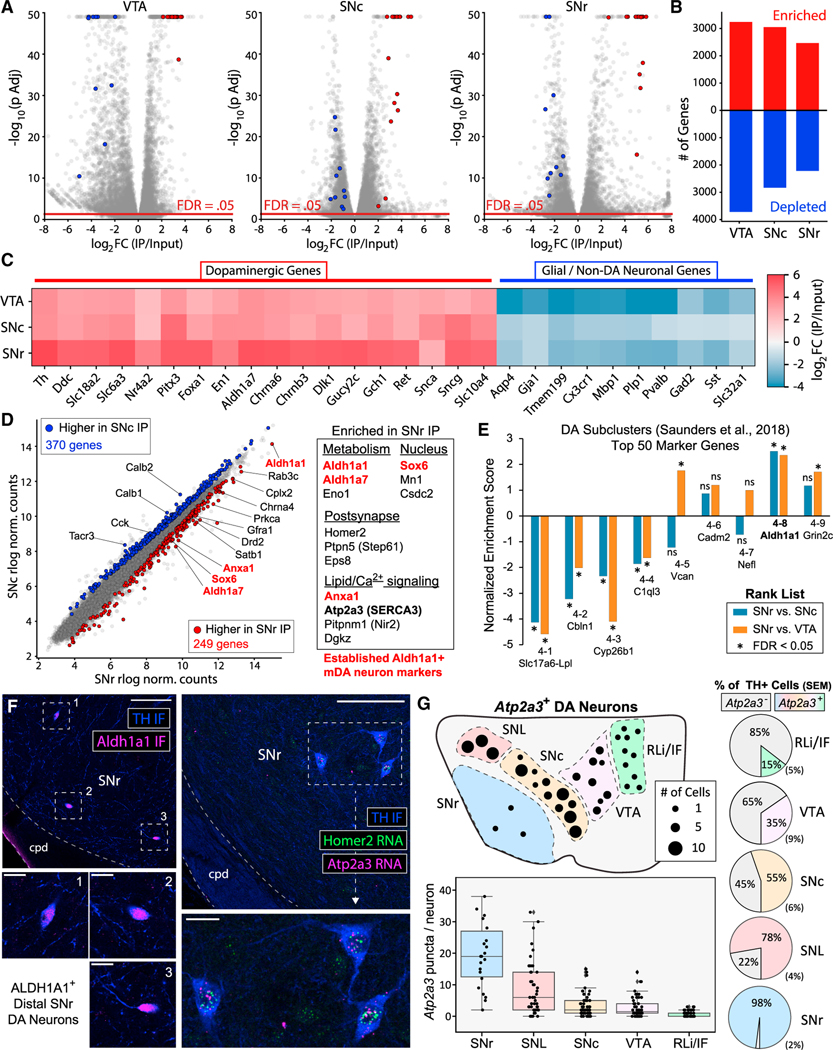
To characterize the mDA neuronal translatome in our SNr dissections (Figure 2), we conducted full-length total RNA-seq (see STAR Methods) of Input and RiboTag IP samples from the SNc, VTA, and SNr. Comparison of IP versus Input samples from Cre+ mice revealed a core enrichment signature of canonical dopaminergic genes (e.g., Th, Ddc, Slc18a2/VMAT2, Slc6a3/ DAT, Pitx3, En1, Ret, and Gch1) in all three regions (Figures 5A–5C). PCA clearly separated IP versus Input samples along PC1 and Regions along PC2 (Figure S5A). The enrichment factor for genes expected to be highly enriched in mDA neurons (Brichta et al., 2015; Saunders et al., 2018) was strikingly higher in SNr IPs than in VTA and SNc IPs (Figure 5C). Together with the low yield from SNr samples (Figure 2), these results suggested that tagged ribosomes derived from a few SNr mDA neurons dominated the RNA content of our SNr RiboTag IPs. Twenty marker genes of various mDA neuronal subpopulations (Poulin et al., 2020) were sufficient for accurate clustering of RiboTag IP samples from all three regions (Figures S5B and S5C). These data demonstrate that many mDA neuronal subpopulation markers identified at the embryonic or early postnatal timepoints are still differentially translated in adult mice. The key markers of the Aldh1a1+/Sox6+ ventral-tier SNc population, which is particularly vulnerable in models of Parkinson’s disease (PD) (Cai et al., 2014; Liu et al., 2014; Poulin et al., 2014), were enriched in our SNr RiboTag IPs (Aldh1a1, Sox6, Aldh1a7, and Anxa1).
Figure 5. SNr RiboTag translatome reveals Aldh1a1+ molecular signature and Atp2a3 (SERCA3) expression in SNr mDA neurons in adult (10–14 mo) mice.
(A) RiboTag IP versus Input volcano plots for VTA, SNc, and SNr samples (n = 4 each). Red or blue points are specific to mDA neurons or to glia and non-mDA neurons, respectively, and shown in (C).
(B) Number of RiboTag IP-enriched or depleted genes (FDR < 0.05) from (A).
(C) Heatmap of average RiboTag IP enrichment for VTA, SNc, and SNr RiboTag IP (n = 4 each).
(D) Average DESeq2 rlog normalized counts for SNr and SNc RiboTag IP samples (n = 4 each). Red and blue genes are differentially expressed between SNr and SNc IP samples. Genes in red are markers of Aldh1a1+/Sox6+ ventral-tier SNc mDA neurons.
(E) Pre-ranked GSEA for SNr versus SNc/VTA IP rank lists using the top 50 marker genes of mDA neuronal clusters from Saunders et al. (2018) as gene sets. *FDR < 0.05 for a rank list/gene set combination.
(F) (Left) TH and ALDH1A1 IF. Scale bars, 100 μm (main) and 20 μm (insets). (Right) FISH for Atp2a3 (SERCA3) and Homer2 in the SNr. Scale bars, 100 μm (main) and 20 μm (inset).
(G) Quantification of Atp2a3 FISH. (Upper left) Anatomical representation of TH+/Atp2a3+ neurons in a single hemi-section (approximately 15 μm thick) containing the indicated VM regions (−3.2 μm posterior to Bregma). Each dot represents 1, 5, or 10 mDA neurons approximating the average of 5 hemi-sections from 4 mice: (RLi/IF) 9.8, (VTA) 31.6, (SNc) 62.8, (SNL) 28.8, and (SNr) 2.8. (Right) Average TH+/Atp2a3− or TH+/Atp2a3+ mDA neurons ±SEM as indicated, corresponding to the upper left. Total cell counts for each region (TH+ neurons/TH+Atp2a3+ neurons): RLi/IF (49/338), VTA (158/503), SNc (314/555), SNL (144/187), SNr (28/29). (Lower left) Boxplot of Atp2a3 mRNA puncta per TH + neuron in the indicated regions from 5 sections, 4 mice per region: (SNr) 22 cells, (SNL) 47 cells, (SNc) 64 cells, (VTA) 58 cells, and (RLi/IF) 58 cells.
We identified mRNAs differentially expressed between SNr and SNc IPs (Figure 5D), with further filtering to select only RiboTag enriched genes (see STAR Methods). We identified 249 genes with greater abundance in SNr versus SNc IPs, including the ventral-tier SNc mDA neuronal markers noted above (Aldh1a1, Sox6, Aldh1a7, and Anxa1) (Figure 5D) (Data S6). Other SNr-enriched mRNAs encoded proteins involved in lipid/calcium signaling, metabolism, and postsynaptic function (Figures 5D, S5F–S5H). Consistent with an enrichment of Aldh1a1+/Sox6+ mDA neurons in our SNr dissection, gene set enrichment analysis (GSEA) (Subramanian et al., 2005) revealed that the top 50 marker genes of the Aldh1a1+ cluster in Saunders et al. (2018) were significantly enriched in SNr versus SNc/VTA IPs (Figure 5E). All of the few TH+ mDA neurons within the SNr were ALDH1A1+ (Figures 5F and S6A). We used FISH to study the distribution of SNr-enriched mRNAs that were not previously described as markers of Aldh1a1+/Sox6+ mDA neurons (Atp2a3, Homer2, Dgkz, and Prkca). These mRNAs were present within mDA neuronal somata in the SNr, but we found no evidence of dendritic localization (Figure 5F, S6B–S6D). Thus, our SNr RiboTag IP predominantly reflects the translatome of SNr mDA neurons, and a molecular signature consistent with Aldh1a1+/Sox6+ mDA neurons in the ventral-tier SNc (Poulin et al., 2020; Saunders et al., 2018). These data are consistent with earlier interpretations that SNr mDA neurons are displaced SNc mDA neurons (Guyenet and Crane, 1981; van der Kooy, 1979) with electrophysiological properties similar to SNc mDA neurons (Brown et al., 2009; Richards et al., 1997).
The SNr enrichment of Atp2a3 mRNA encoding the sarco/ER Ca2+-ATPase isoform 3 (SERCA3) is of particular interest given the importance of cytosolic Ca2+ oscillations in mDA neurons (Zampese and Surmeier, 2020). SERCA3 is predominantly expressed in hematopoietic and endothelial cells (Bobe et al., 1994; Burk et al., 1989; Wuytack et al., 1994) and has an approximately 5-fold lower affinity for Ca2+ than the ubiquitous SERCA2 isoform (Lytton et al., 1992). Our RiboTag data indicate that mDA neuronal translation of Atp2a3/SERCA3 in all VM regions, although relative abundance was greatest in the SNr (Figure 5E). We used FISH to define the anatomical distribution of Atp2a3/ SERCA3-expressing mDA neurons. In addition to the SNr (Figure 5F), we found Atp2a3/SERCA3+ mDA neurons in the substantia nigra pars lateralis (SNL), SNc, VTA, and midline nuclei (rostral linear nucleus and interfascicular nucleus [RLi/IF]) (Figures S6E–S6H). While few in number, nearly all SNr mDA neurons were Atp2a3/SERCA3+ and expressed the highest mRNA levels per neuron (Figure 5G). mDA neurons in the SNL also express high levels of Atp2a3/SERCA3, while Atp2a3/SERCA3 expression was extremely sparse in the midline nuclei (Figure 5G). Cerebellar Purkinje neurons also express SERCA3 (Baba-Aïssa et al., 1996) and exhibit pacemaker firing (Raman and Bean, 1999). Given that SERCA3 expression is altered in PD (Aguila et al., 2021) and may regulate cytosolic Ca2+ dynamics, future studies should investigate SERCA3 function in mDA neurons.
Midbrain synaptosome RiboTag IP reveals dendritic localization of mRNAs encoding vesicular release proteins
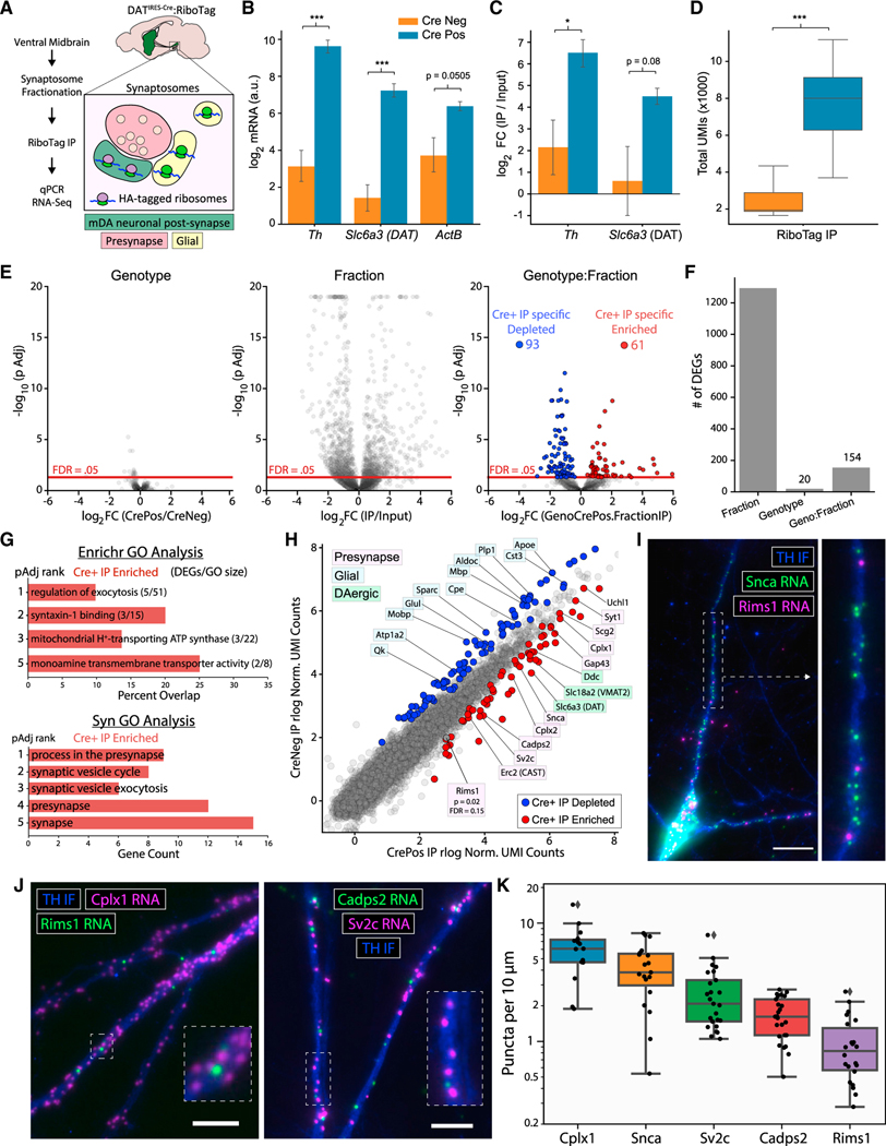
Another approach to identify translating mRNAs in dendrites is to combine cell type-specific ribosome IP with subcellular fractionation (Ouwenga et al., 2017, Ouwenga et al., 2018). SNc mDA neurons possess dendritic spines (Hage and Khaliq, 2015; Jang et al., 2015), and resealed dendritic elements within the midbrain synaptosome preparations exhibit DA release and reuptake (Hefti and Lichtensteiger, 1978a, 1978b; Silbergeld and Walters, 1979). We conducted RiboTag IP on synaptosomes prepared from VM tissue (Figure 6A). qRT-PCR revealed greater yield of Th, Slc6a3/DAT, and Actb mRNA in Cre+ IPs compared with Cre− controls (Figure 6B). Similarly, Th and Slc6a3/DAT enrichment was approximately 16-fold greater in Cre+ IP versus Input comparisons compared with Cre− controls (Figure 6C). Given the absence of local axon collaterals from mDA neurons (Omelchenko and Sesack, 2009; Tepper et al., 1987), these data demonstrate mDA neuron-specific ribosome capture from dendritic or postsynaptic elements.
Figure 6. Dendritic localization of mRNAs encoding exocytosis and vesicular release proteins in DA neurons.
Data in Figures 6A–6H are from adult mice (10–14 mo); Figures 6I–6K are from postnatal mDA neuronal cultures.
(A) Schematic for midbrain synaptosome fractionation and RiboTag IP.
(B) qRT-PCR of Th, Slc6a3/DAT, and Actb from RiboTag IP of VM synaptosomes from Cre− (n = 5) and Cre+ mice (n = 6). Log2 mRNA abundance is in arbitrary units (40 – Cq). Two-way ANOVA main effects for genotype, F = 98.2 (1, 27), p = 1.7e-10; RNA, F = 6.10 (2, 27), p = 0.0065; and genotype:RNA interaction, F = 5.47 (2, 27), p = 0.010. Tukey’s HSD post hoc test for Cre− versus Cre+ IP samples (p-adj < 0.001 for Th and DAT, p-adj = 0.0505 for ActB). ***p < 0.001.
(C) qRT-PCR for RiboTag IP/Input enrichment relative to Actb (mean ΔΔCq +/− SEM), n = 5 Cre− and n = 6 Cre+ mice. *p < 0.05, Welch’s t test.
(D) UMIs per sample for VM synaptosome RiboTag IPs from Cre− (n = 6) and Cre+ mice (n = 8). ***p < 0.001, Welch’s t test.
(E) Volcano plots from DESeq2 with GLM: ~genotype + fraction + genotype:fraction. (Left) Genotype effect. (Middle) fraction effect. (Right) Genotype:fraction interaction. log2(GenoCrePos.FractionIP) is the difference in the fraction effect between genotypes: {Cre+ log2FC(IP/Input) – Cre− log2FC(IP/Input)}.
(F) Number of DEGs (FDR < 0.05) from (E).
(G) GO analysis (upper: Enrichr; lower, SynGO) of Cre+ IP enriched genes from (E).
(H) Average DESeq2 rlog normalized UMI counts for Cre+ (n = 8) and Cre− (n = 6) VM synaptosome IPs. Red/blue genes indicate Cre+ IP enriched/depleted genes (FDR < 0.05). Genes with dopaminergic (green), glial (blue), and presynaptic function (pink) are labeled.
(I) TH IF and FISH (RNAscope) for Snca and Rims1 mRNA in cultured mDA neurons. Dashed white lines indicate the inset (right). Scale bar, 20 μm.
(J) TH IF and FISH for Cplx1 and Rims1 (left) or Cadps2 and Sv2c mRNA (right) in the dendrites of cultured mDA neurons. Dashed white lines indicate inset (right). Scale bars, 10 μm. (K) Quantification of RNA puncta from (I–J) per 10 μm of dendrite. Data are from 2–3 independent cultures, with n dendrites quantified for each mRNA: Cplx1 (n = 16), Snca (n = 19), Sv2c (n = 25), and Cadps2 (n = 25), Rims1 (n = 35).
We next analyzed the mDA neuronal translatome of midbrain synaptosome samples using 3′ UMI-based RNA-seq. Consistent with the increased mRNA yield measured by qRT-PCR, Cre+ IP samples had significantly more UMIs than Cre− controls (Figure 6D). As in the striatum, we found a significant effect of fraction for more than 1,000 genes (Figures 6E and 6F), reflecting genotype-independent non-specific binding. However, in contrast with all striatal RiboTag IP experiments, we observed a significant effect of genotype:fraction interaction for 154 genes (Figures 6E and 6F). These genes are significantly depleted (93) or enriched (61) in IP compared with Input samples only in Cre+ mice (see Data S7). Similar to striatal synaptosomes, glial mRNAs such as Apoe, Cst3, Cpe, Glul, Mbp, and Plp1 were abundant in midbrain synaptosomes; however, these glial mRNAs were uniformly depleted from Cre+ IP samples (Figure 6H). Strikingly, GO analysis of the Cre+ IP-enriched genes revealed a significant overrepresentation of terms such as regulation of exocytosis, process in the presynapse, and synaptic vesicle exocytosis (Figure 6G). Thus, in addition to canonical dopaminergic mRNAs, we found Cre-dependent enrichment of mRNAs encoding a wide range of proteins with presynaptic function (Figure 6H). These include mRNAs encoding proteins involved in synaptic vesicle fusion and recycling (Erc2/CAST, Cplx1, Cplx2, Syt1, Sv2c, and Snca) and dense core vesicle release (Cadps2/CAPS2 and Scg2) (Figure 6H). We also observed a near-significant enrichment of Rims1, which encodes the active zone protein RIM1 that is involved in both axonal and somatodendritic DA release (Liu et al., 2018; Robinson et al., 2019). Many of these mRNAs have also been identified in the dendrites of both glutamatergic and GABAergic hippocampal neurons (Perez et al., 2021) (Figure S7A).
We validated the dendritic localization of several mRNAs encoding release proteins in mDA neuron cultures (Rayport et al., 1992). We first confirmed that cultured mDA neurons recapitulate the dendritic localization of Th, Ddc, Slc6a3/DAT, and Slc18a2/VMAT2 mRNAs observed in the VM (Figures S7B–S7E). Although α-synuclein is abundant in presynaptic varicosities, we found dense localization of Snca mRNA in dendrites (Figure 6I). Similarly, we found a striking density of Cplx1 (Complexin 1) mRNA in dendrites, along with scattered Rims1 mRNA (Figure 6J). The Ca2+-dependent activator protein of secretion 2 (CAPS2) is involved in catecholamine loading into dense core vesicles in neuroendocrine cells (Brunk et al., 2009; Ratai et al., 2019) and is particularly enriched in mDA neurons (Sadakata et al., 2006). We found Cadps2/CAPS2 mRNA within dopaminergic dendrites, along with Sv2c mRNA (Figure 6J). Synaptic vesicle glycoprotein 2C (SV2C) is involved in axonal DA release (Dunn et al., 2017) and may also play a role in somatodendritic DA release. Consistent with the midbrain synaptosome IP (Figure 6H), quantification of mRNA puncta revealed that Cplx1 and Snca were most abundant in dendrites, followed by Sv2c and Cadps2, and Rims1 (Figure 6K). Collectively, these data suggest that the local translation of vesicular release proteins may regulate dendritic DA release in mDA neurons.
DISCUSSION
Predominance of somatic translation in mDA neurons
We used multiple approaches to characterize the subcellular distribution of tagged ribosomes in mDA neurons, each of which identified the soma as the major site of protein synthesis. The absence of axonal mRNA localization by FISH (Figure 4) and lack of mRNA capture by striatal RiboTag IP (Figure 3) supports the absence of translating ribosomes in mDA axons. The paucity of eL22-HA in mDA axons is surprising, given their massive axonal arborization. Strikingly, while the striatal axons of SNc mDA neurons likely comprise more than 90%–95% of their cellular volume (e.g., Matsuda et al., 2009) and approximately 90% of their cellular protein (Hobson et al., 2021), we found only approximately 1% of eL22-HA in the striatum (Figure 2D). If all of this eL22-HA were present in functional ribosomes, the axonal ribosome/protein ratio would be 103–104 lower than in mDA neuronal perikarya. Our results suggest that mDA striatal axons are supplied by a combination of slow and fast axonal transport (Maday et al., 2014; Roy, 2014) of somatically synthesized proteins. Indeed, the massive bioenergetic burden placed on axonal transport systems in mDA neurons may contribute to their demise in PD (Chu et al., 2012; Sulzer, 2007).
Dopaminergic mRNA localization and translation in dendrites
In addition to their massive axonal arbors, SNc mDA neurons must supply SNr dendrites with machinery for DA synthesis, release, and reuptake. Here, we show that Th, Ddc, Slc6a3/DAT, and Slc18a2/VMAT2 mRNAs are localized throughout mDA neuronal dendrites in the SNr (Figure 4) and are bound to dopaminergic ribosomes in midbrain synaptosomes (Figure 6). In conjunction with vesicular sorting mechanisms (Li et al., 2005), dendritic translation could rapidly modify the local abundance of DA transmission machinery. DAT is often localized on vesicular and tubular membrane elements within dendrites (Hersch et al., 1997; Nirenberg et al., 1996a). VMAT2 is also found on similar structures, termed tubulovesicles, that seem to consist of smooth ER and may represent the site of dendritic DA storage and release (Cheramy et al., 1981; Nirenberg et al., 1996b). The local synthesis of DAT and VMAT2 would require the presence of dendritic ER and a local secretory pathway. Although mDA neuronal dendrites are devoid of obvious Golgi apparatus in ultrastructural studies (Nirenberg et al., 1996b), local processing could occur in Golgi outposts (Hanus and Ehlers, 2016) or recycling endosomes (Bowen et al., 2017). The tubulovesicles could also be related to ribosome-associated vesicles, a novel ER subcompartment found in secretory cells and neuronal dendrites (Carter et al., 2020). Thus, the translation of DAT and VMAT2 within local secretory pathways may be linked to the biogenesis of DA storage and release structures within mDA dendrites.
Beyond the core dopaminergic machinery, how do mDA neurons manage simultaneous axonal and dendritic localization of vesicular release proteins? For proteins involved in both axonal and somatodendritic DA release, such as RIM1 (Liu et al., 2018; Robinson et al., 2019), our data show that protein trafficking supplies the vast majority of protein to striatal mDA axons. In contrast, the local translation of RIM1 and other release proteins such as complexins may be important for establishing exocytic release sites in dopaminergic dendrites (Figure 6). Postsynaptic complexins are known to regulate AMPA receptor exocytic events during long-term potentiation (Ahmad et al., 2012) and could be involved in dendritic DA release.
The molecular characteristics of the organelles and fusion mechanisms that mediate somatodendritic DA release remain unclear (Rice and Patel, 2015). When expressed in hippocampal neurons, VMAT2 colocalizes with brain-derived neurotrophic factor on vesicles that undergo regulated exocytosis in dendrites (Li et al., 2005). Given that CAPS2 regulates the release of neurotrophin-containing vesicles in cerebellar granule cells (Sadakata et al., 2004) and co-localizes with SNAP-25 in mDA neuronal soma (Sadakata et al., 2006), the dendritic translation of Cadps2/CAPS2 mRNA (Figure 6) raises the possibility of CAPS2 involvement in dendritic DA release. More broadly, it is possible that the trafficking of synaptic vesicle release proteins in mDA neurons has been optimized to shuttle them into striatal axons, and that such a polarization is incompatible with simultaneous trafficking into dendrites. Local translation in dopaminergic dendrites may provide an alternative mechanism of localization for these proteins, enabling the dynamic regulation of proteins at the precise intracellular sites of dendritic exocytosis.
Limitations of the study
While our findings are supported by multiple, independent approaches, there are limitations. We cannot definitively exclude the possibility that extremely low levels of axonal ribosomes are present below our limit of detection, since the visualization of eL22-HA in retinal axons required immunoelectron microscopy (Shigeoka et al., 2016). Tagged ribosomal proteins did not prevent the axonal localization of ribosomes in other neurons (Ostroff et al., 2019; Shigeoka et al., 2016), but we also cannot rule out that the HA-tag in eL22 somehow interferes with axonal localization in mDA neurons. However, we were able to reliably detect low levels of eL22-HA on translating ribosomes in mDA neuronal dendrites. We found that the presence of only a few mDA neuronal soma in the SNr dominated dendritic ribosomes in RiboTag IPs from SNr dissections (Figure 5); future studies of dendritic DA release in the SNr should be designed to avoid SNr mDA neurons. In our synaptosome RiboTag IP studies of the dendritic translatome, we confirmed the dendritic localization of key candidate mRNAs using FISH (Figure 6). Nonetheless, it is possible that some mDA neuronal ribosomes in the synaptosome fraction are derived from non-synaptic cellular elements. Future work on subcellular mRNA localization may leverage super-resolution imaging (Alon et al., 2021; Eng et al., 2019; Wang et al., 2020).
STAR★METHODS
Detailed methods are provided in the online version of this paper and include the following:
RESOURCE AVAILABILITY
Lead contact
Further information and requests for resources should be directed to and will be fulfilled by the Lead Contact, Peter Sims (pas2182@columbia.edu).
Materials availability
This study did not generate new unique reagents.
Data and code availability
The RNA-seq data generated in this study are publicly available on the NIH Gene Expression Omnibus database (GEO: GSE180913). Raw count matrices and differential expression analysis output are provided as supplemental information.
The Python and Shell code used for processing of RNA-seq data is accessible at: https://github.com/simslab/DropSeqPipeline8 (https://doi.org/10.5281/zenodo.5534458), and Python code for FISH analysis is accessible at: https://github.com/simslab/Neurite_FISH_Quant (https://doi.org/10.5281/zenodo.5570748).
Any additional information required to reanalyze the data reported in this paper is available from the lead contact upon request.
EXPERIMENTAL MODEL AND SUBJECT DETAILS
Animals
All animals were housed in a 12-h/12h light/dark cycle with ad libitum access to food and water. DATIRES-Cre mice (JAX #006660, RRID: IMSR_JAX:006660) (Bäckman et al., 2006), Ai9 mice (JAX #007909, RRID: IMSR_JAX:007909) (Madisen et al., 2010) and RiboTag mice (JAX #029977, RRID: IMSR_JAX:029977) (Sanz et al., 2009) were obtained from Jackson Laboratories. DAT-Cre mice (MGI:3770172, RRID: MGI:3770172) (Turiault et al., 2007) used in the FASS studies were a kind gift from Dr François Tronche. VGLUT1VENUS mice (Slc17a7tm1.1Ehzg, RRID: 5297706) used in the FASS studies have been previously described (Biesemann et al., 2014; Herzog et al., 2011).
Middle aged adult mice (10–14 months of age) of both sexes were used in most experiments unless otherwise noted, except for DA FASS studies, which used mature adult mice (3–6 months) of both sexes. For RiboTag experiments involving early postnatal ages (P0–P31), mice of both sexes were used and the exact ages are indicated in the text and figure captions. DATIRES-Cre:RiboTag experimental litters were bred by crossing homozygous RiboTag mice (RiboTag+/+) with heterozygous DATIRES-Cre (DATIRES-Cre/wt) mice, yielding litters of DATIRES-Cre/wt;RiboTag+/− (Cre-positive) and DATwt/wt;RiboTag+/− (Cre-negative) mice. Experimenters were blind to the genotype of mice in these litters throughout animal sacrifice and tissue dissection. Genotyping for the DATIRES-Cre allele was conducted before biochemical experiments using established protocols (Bäckman et al., 2006). All experimental procedures were conducted according to National Institutes of Health guidelines and were approved by the Institutional Animal Care and Use Committees of Columbia University and the New York State Psychiatric Institute, or according to the European guide for the care and use of laboratory animals and approved by the ethics committee of Bordeaux Universities (CE50) under the APAFIS #21132–2019061314534378v4 (CNRS, France).
METHOD DETAILS
Viral injections
As previously described (Paget-Blanc et al., 2021), Stereotaxic injections were performed in heterozygous DAT-Cre+ mice of either sex at 8–9 weeks of age. An AAV1 pCAG-FLEX-EGFP-WPRE from the University of Pennsylvania core facility (Oh et al., 2014) was injected into DAT-Cre+ mice. Saline-injected littermates were used as autofluorescence controls. The stereotaxic injections were performed in isoflurane-anesthetized mice using a 30-μL glass micropipette. Injection coordinates for the SNc were anterior/posterior (A/P), 3.6 mM; lateral (L), ±1.3 mM; and dorsal/ventral (D/V), 4.2 mM. Injection coordinates for the VTA were A/P, 3.16 mM, L, ±0.6 mM; and D/V, 4.2 mM. The A/P and L coordinates are with respect to the bregma, whereas the D/V coordinates are given with respect to the brain surface. The animals were euthanized after 28 days at the maximal viral EGFP expression. For FASS experiments, four to six DAT-Cre+ mice and one wild-type mouse were used.
| Antibody use | |
|---|---|
|
| |
| Name | Use |
| Rabbit anti-HA | IHC, 1:1000 |
| Chicken anti-TH | IHC, 1:500 |
| ICC, 1:1000 | |
| Rabbit anti-RFP | IHC, 1:500 |
| Rabbit anti-ALDH1A1 | IHC, 1:500 |
| Goat anti-chicken IgY (H+L), Alexa Fluor Plus 488 | IHC/ICC, 1:1000 |
| Goat anti-rabbit IgG (H+L), Alexa Fluor 647 | IHC/ICC, 1:1000 |
| Goat anti-rabbit IgG HRP | IHC, 3:10,000 |
| Mouse anti-HA | WB, 1:1000 |
| Goat anti-mouse HRP | WB, 1:5000 |
See the Key resources table for manufacturer/catalog numbers. IHC, staining of acute brain slices or sections; ICC, staining of cultured neurons; WB, Western blotting.
Neuronal cultures
Ventral mesencephalic cultures containing dopaminergic neurons were prepared according to established procedures (Rayport et al., 1992). The VM (SN and VTA) from postnatal days 0–2 mice of either sex was dissected, dissociated, and plated on a monolayer of rat cortical astrocytes at the plating density of approximately 100,000 cells/cm2. Experiments were conducted 14–21 d after plating.
Immunohistochemistry
Mice were anesthetized with euthasol and transcardially perfused with approximately 15 mL of 0.9% saline followed by 40–50 mL of ice-cold 4% paraformaldehyde (PFA) in 0.1 M phosphate buffer (PB), pH 7.4. Brains were post-fixed in 4% PFA in 0.1M PB for 6–12 h at 4°C, washed three times in phosphate-buffered saline (PBS), and sectioned at 50 μm on a Leica VT1000S vibratome. Sections were placed in cryoprotectant solution (30% ethylene glycol, 30% glycerol, 0.1M PB, pH 7.4) and stored at −20°C until further use.
Sections were removed from cryoprotectant solution and washed three times in tris-buffered saline (TBS) at room temperature. Sections were then permeabilized in TBS + 0.2% Triton-X 100 for 1 h at room temperature, followed by blocking in TBS + 10% normal goat serum (NGS) and 0.3% Triton-X 100 for 1.5 h at room temperature. Sections were then directly transferred to a prechilled solution containing primary antibodies in TBS + 2% NGS + 0.1% Triton-X 100 and incubated for approximately 40 h at 4°C. Sections were washed in TBS + 0.05% Tween 20 (TBS+T) five times over 1 h at room temperature. Sections undergoing tyramide signal amplification were treated with 3% hydrogen peroxide in TBS + T for 15 min at room temperature, followed by another two washes in TBS + T. Sections were incubated in a solution containing secondary antibodies in TBS + 2% NGS + 0.1% Triton-X 100 at room temperature for 1.5 h, followed by four washes in TBS + T over 45 min at room temperature. Sections undergoing tyramide signal amplification were then incubated in TSA-Cy5 (Perkin Elmer; 1:7500) in the manufacturer’s diluent buffer for 1 h at room temperature. After four additional washes in TBS, sections were slide mounted and coverslipped with Fluoromount G (Southern Biotech). See Antibody use for a complete list of antibodies and concentrations used in this study.
Tissue dissection for RiboTag IP
Mice were sacrificed by cervical dislocation and brains were rapidly extracted and submerged in ice-cold 0.32 M sucrose buffer with 5 μm HEPES pH 7.4, 10 μm MgCl2, and 100 mg/mL cycloheximide (CHX). Brains were placed on an ice-cold brain matrix (Zivic Instruments) and separated into 0.5- to 1.0-mm sections using ice cold razor blades. Striatum was dissected from slices between approximately –0.5 mm and 1.5 mm AP to Bregma. To avoid potential DATIRES-Cre recombined cells in the lateral septum, a single vertical cut was made descending from the lateral ventricle on each side, and all medial tissue (including the lateral septum and nucleus accumbens shell) was discarded. The corpus callosum, cortex, and ventral olfactory tubercle were removed. The remaining dorsal and ventral striatum tissue was flash frozen on liquid nitrogen and stored at −80°C.
VM tissue was dissected from slices between approximately −2.5 mm and −3.75 mm AP to Bregma. First, the cortex, hippocampi, and any hypothalamus or white matter ventral to the midbrain were removed. For whole VM tissue dissections, a single horizontal cut was made just dorsal to the rostral linear nucleus and all dorsal tissue was discarded. The remaining tissue containing the SN/VTA was flash frozen on liquid nitrogen and stored at −80°C. For regional dissections, the SNr was first dissected away from the midbrain using a conservative semilunar cut halfway from the edge of the cerebral peduncle to the SNc (see Figure 2A). The remaining SNc tissue on either side was separated from the VTA by a vertical cut at the lateral edge of the VTA. All tissues were flash frozen and stored at −80°C.
Synaptosome preparation for RiboTag IP
VM or striatal dissections were homogenized in 1 mL of ice-cold 0.32 M sucrose with 5 mM HEPES pH 7.4, 10 mM MgCl2, 100 μg/mL CHX, 13 EDTA-free protease inhibitors (Roche), and 100 U/mL SUPERaseIN. Nuclei and large debris were cleared at 2,000×g for 10 min at 4°C. The supernatant (S1) was further centrifuged at 7,000×g for 15 min at 4°C to yield the P2 pellet. The supernatant (S2) (cytoplasm and light membranes) was removed from the P2 pellet, which was washed by resuspension in 1 mL of ice-cold 0.32 M sucrose buffer (HEPES, MgCl2, CHX, and inhibitors as above) and re-centrifuged at 10,000×g at 4°C before lysis. P2 pellets were lysed in 1 mL of lysis buffer (5 mM HEPES pH 7.4, 150 mM KCl, 10 μm MgCl2, 1% Igepal CA-620, 100 μg/mL CHX, 1 × EDTA-free protease inhibitors [Roche], and 100 U/mL SUPERaseIN). After resuspension, samples were incubated at 4°C on a rotor for 15 min. The resulting synaptosome lysate was subjected to RiboTag IP as described below.
Fluorescence-activated synaptosome sorting
Synaptosomes were prepared from the striatum or cortex of VGLUT1venus or DAT-Cre eGFP-expressing mice by homogenization in 1 mL of ice-cold isosmolar buffer (0.32 M sucrose, 4 mM HEPES pH7.4, protease inhibitor cocktail Set 3 EDTA-free [EMD Millipore Corp.]), using a 2 mL glass Teflon homogenizer with 12 strokes at 900 rpm. The homogenizer was rinsed with 250 μL of isosmolar buffer and three manual strokes and then, the pestle was rinsed with additional 250 μL of isosmolar buffer. The final 1.5 mL of homogenate (H) was centrifuged at 1000×g for 5 min at 4°C in a benchtop microcentrifuge. The supernatant (S1) was separated from the pellet (P1) and centrifuged at 12,600×g for 8 min at 4°C. The supernatant (S2) was discarded and the synaptosomes-enriched pellet (P2) was resuspended in 0.5 mL of isosmolar buffer and layered on a two-step Ficoll density gradient (900 μL of 7.5% and 900 mL of 13% Ficoll, 4 mM HEPES). The gradient was centrifuged at 50,000×g for 21 min at 4°C (Beckman Coulter Optima MAX XP ultracentrifuge with a TL-55 rotor). Sucrose synaptosomes were recovered at the 7.5/13% Ficoll interface using a 0.5-mL syringe.
Ficoll gradient-purified synaptosomes were diluted in PBS containing 1 μg /mL FM4–64 and stored on ice throughout the FASS procedures. The FACSAria-II (BD Biosciences) was operated with the following settings: 70 μm nozzle, sample shaking 300 rpm at 4°C, FSC neutral density filter 1.0, 488 nm laser on, area scaling 1.18, window extension 0.5, sort precision 0–16-0, FSC (340 V), SSC (488/10 nm, 365V), fluorescein isothiocyanta (enhanced green fluorescent protein) (530/30 nm, 700 V), PerCP (FM4–64) (675/20 nm, 700 V). Thresholding on FM4–64 was set with a detection threshold at 800. Samples were analyzed and sorted at rates of 15,000–20,000 events/s and flow rate of 3. Data were acquired using BD FACS DIVA 6. Cytometry plots were generated using FCS Express 7 (De Novo Software).
FISH
For mouse brain tissue and neuronal cultures, FISH was performed using the highly sensitive RNAScope Multiplex Fluorescent v2 assay (ACD Bio). See Antibodies and Reagents for a complete list of probes and reagents used in this study. Although most single FISH puncta using this assay are likely single mRNA molecules (Wang et al., 2012), this cannot be definitively determined owing to the enzymatic signal amplification and non-diffraction-limited size of the mRNA puncta.
Mouse brain sections were prepared as above, removed from cryoprotectant solution, and washed three times in TBS at room temperature. Sections were incubated with hydrogen peroxide (ACD) for 15 min at room temperature, washed several times in TBS, and then mounted to Superfrost slides (Fisher). Sections were allowed to dry for 10 min and a hydrophobic barrier (PAP pen, Vector Labs) was created around the tissue. Tissue was incubated in 50% EtOH, then 70% EtOH, then 100% EtOH for 5 min each. Sections were rehydrated in TBS for several minutes, digested with Protease IV (ACD) for 25 min at room temperature, and rinsed twice with TBS before proceeding to the RNA Scope Multiplex Fluorescent v2 assay (ACD).
Neuronal cultures were fixed in 4% PFA in 0.1 M PB + 4% sucrose for 10 min at room temperature. After several washes in TBS, the dish was filled with methanol pre-chilled to −20°C. Cultures were stored at −20°C for up to 4 weeks before FISH. After allowing cultures to come to room temperature, methanol was replaced with 70% EtOH at room temperature for 2 min, then with 50% EtOH for 2 min, and then cultures were washed for 10 min in TBS. Cultures were treated with hydrogen peroxide (ACD) for 10 min at room temperature, followed by Protease III (ACD) diluted 1:15 in TBS for 10 min at room temperature, followed by two rinses in TBS before proceeding to the RNA Scope Multiplex Fluorescent v2 assay.
The RNA Scope Multiplex Fluorescent v2 assay was conducted according to the manufacturer’s instructions, with all incubations taking place in a humidified chamber at 40°C. Two 5-min washes in excess RNA Scope Wash Buffer (ACD) took place between each incubation in sequential order: probes (2 h), AMP1 (30 min), AMP2 (30 min), AMP3 (15 min), HRP-C1/2/3 (15 min), TSA Cy3 (1:1500, 30 min), HRP blocker (30 min), HRP-C1/2/3 (15 min), and TSA Cy5 (1:1500, 30 min). Samples were washed twice more in RNA Scope Wash Buffer, then twice more in TBS. Samples were then blocked and immunostained for TH as described above. After immunostaining, samples were mounted in Fluoromount G and stored at 4°C for up to 1 week before imaging.
RiboTag ribosome IP
A detailed protocol is available online at protocols.io (https://doi.org/10.17504/protocols.io.by37pyrn): https://doi.org/10.17504/protocols.io.by37pyrn.
Frozen tissues were thawed on ice in a glass-glass dounce homogenizer with 1–1.5 mL of ice-cold lysis buffer (20 μm HEPES pH 7.4, 150 mM KCl, 10 mM MgCl2, 0.5 mM DTT, 100 μg/mL CHX, 1× EDTA-free protease inhibitors [Roche], and 100 U/mL SUPERaseIN). Tissues were lysed on ice using 30 strokes each with A and B pestles. Lysates were transferred to pre-chilled Eppendorf tubes and centrifuged at 1,000×g 4°C for 10 min, after which the supernatant was transferred to a new tube. One-ninth of the volume of 10% Igepal CA-630 was added to the lysates (final concentration 1%) and they were rotated at 4°C for 15 min. Lysates were clarified by centrifuging at 20,000×g 4°C for 10 min and transferred to a new tube. Five percent of the lysate was reserved as Input and frozen at −80°C.
We then added 1.5 μg (for striatal samples) or 6 μg (for midbrain samples) of biotinylated rabbit anti-HA and the lysates were rotated overnight at 4°C. Compared with previous protocols using Protein G Dynabeads, we found that biotinylated anti-HA IgG and streptavidin T1 Dynabeads enabled rapid binding with higher specificity (Figure S1H). Streptavidin T1 Dynabeads (ThermoFisher, catalog #65601) were then added to the lysates (5 μL/μg of biotinylated antibody) and rotated for 30 min at 4°C. Beads were captured on a magnetic rack and the lysate was discarded. Beads were resuspended in 500 μL of ice-cold high salt buffer (20 mM HEPES, pH 7.4, 350 mM KCl, 10 mM MgCl2, 1% Igepal CA-630, 0.5 mM DTT, 100 μg/mL CHX, 1× EDTA-free protease inhibitors [Roche], and 100 U/mL SUPERaseIN) and transferred to a new tube. Beads were rotated for 30 min at 4°C, then captured on a magnetic rack and washed again three more times with ice-cold high salt buffer (four washes total over 2 h). After the last wash, beads were resuspended in 100 μL of ribosome release buffer (20 μm HEPES, pH 7.4, 50 μm EDTA, 100 U/mL SUPERaseIN) and incubated for 10 min at room temperature. Beads were captured on a magnetic rack and the eluate containing the released mRNA was transferred to a new tube. Beads (with eL22-HA still bound) were flash frozen on liquid nitrogen and stored at −80°C. The aqueous mRNA eluate was purified using the RNEasy MinElute kit (Qiagen, catalog #74204) according to the manufacturer’s instructions. RNA was eluted in 14 μL of nuclease free water supplemented with 20 U/mL SUPERaseIN and stored at −80°C.
Quantitative RT-PCR
For RiboTag IP samples, an equal fraction of captured RNA was reverse transcribed (1.5 μL of the 14 μL elution from RNEasy MinElute purification). For Input or other tissue samples, 20–50 ng of total RNA was reverse transcribed. RNA was reverse transcribed in a 20-μL reaction with 0.5 U of Maxima H Reverse Transcriptase (ThermoFisher, catalog # EP0753) and random hexamers (5 μm, ThermoFisher catalog #SO142).
A quantitative PCR was run with TaqMan Universal Master Mix (ThermoFisher catalog #4440042) and TaqMan FAM-MGB primer/ probe sets spanning exon junctions on a BioRad CFX96. The following primer/probe sets were used (ThermoFisher): Mouse ActB, Μm01205647_g1; Mouse Th, Μm00447557_m1; Mouse Slc6a3/DAT, Μm00438388_m1; Mouse Slc18a2/VMAT2, Μm00553058_m1; Mouse Gfap, Μm01253033_m1; Mouse Mbp, Μm01266402_m1; and ERCC-0096, Ac03460023_a1. For RiboTag IP samples, an equal fraction of cDNA was used in each reaction. For Input or other tissue samples, 3–5 ng cDNA was used in each reaction.
Western blotting
Frozen Streptavidin T1 Dynabeads were thawed and resuspended in 1× LDS sample buffer supplemented with 20 mM DTT. To elute eL22-HA, beads were boiled at 95°C for 5 min and then placed onto a magnetic rack. Samples were loaded into 10% Bis-Tris poly-acrylamide gels (Invitrogen, ThermoFisher catalog #NP0303BOX) and transferred to PVDF membranes (Immobilon-P, Millipore-Sigma, catalog #IPVH00010). Membranes were initially washed for 15 min in TNS with Tween (TBST) (1× TBS + 0.1% Tween 20), blocked for 1 h in 5% bovine serum albumin (BSA)/TBST, and incubated overnight at 4°C with primary antibody in 5% BSA/TBST overnight. After primary incubation, membranes were washed three times in TBST before incubation with horseradish peroxidase (HRP)-conjugated secondary antibody in 5% BSA/TBST for 1 h at room temperature. After secondary incubation, membranes were washed three times in TBST. Signal was developed using Immobilon enhanced chemiluminescent substrate (Millipore, catalog #WBKLS0500) and imaged on an Azure Biosystems C600 system.
Image acquisition
Widefield imaging of eL22-HA staining was conducted on a Nikon Ti2 Eclipse equipped with a SpectraX light engine (Lumencor) and a DS-Qi2 camera (Nikon), using a 10x/0.25 NA or 20x/0.75 NA air objectives (Nikon). Confocal imaging was conducted on a Leica SP8 laser scanning system using a 60×/1.45 NA objective (Leica).
eL22-HA image analysis
Z stack images from the SNr, MFB, and striatum were acquired using a 60×/1.45 NA oil-immersion objective on a Leica SP8 confocal microscope. The 10-μm depth sub-stacks were collapsed via maximum projection for downstream analysis. A binary mask was used to identify pixels in TH-positive dendrites and axons. The mean eL22-HA intensity for TH-positive pixels was subtracted from the mean eL22-HA intensity for all pixels within each field and is reported in Figure 1F as normalized eL22-HA mean fluorescence intensity.
FISH image analysis
RNA puncta were analyzed using TrackMate (Tinevez et al., 2017). The Laplacian of Gaussian spot detector with estimated blob diameter of 0.5–1.0 μm. Additional filtering was implemented using a combination of quality, contrast, and total intensity as necessary to suppress background spot detection. For each image, the centroid (X,Y,Z) coordinates, diameter, and other quantitative parameters of each punctum were exported for further analysis.
For the percent co-localization shown in Figure 4D, a binary threshold was set for the TH IF signal based on two standard deviations above the image background to generate a binary mask of pixels for TH+ neurites. The 23 pixels surrounding the TrackMate centroid coordinate of each punctum were analyzed (3 × 3 × 3 cube of pixels excluding the four corner pixels) for overlap with the TH+ neurite pixels. Puncta with more than 60% overlapping pixels were retained as co-localized within TH+ neurites. The number of puncta co-localized within TH+ neurites was divided by the total volume of TH+ pixels in each field, yielding the puncta per volume of TH+ neurite shown in Figure 4D.
For quantification of puncta per micrometer of dendrite shown in Figure 4F, individual dendrites were segmented using the SimpleNeuriteTracer plugin in ImageJ. Dendrites were filled in three dimensions and exported as a binary mask, from which the (X,Y,Z) coordinates of all pixels in each dendrite were extracted. TrackMate was run once on each original image file, and the number of puncta within each dendrite was determined using the same co-localization analysis as above. The number of puncta in each dendrite was divided by the path length of each dendrite from SimpleNeuriteTracer.
For the quantification of Atp2a3 puncta per neuron shown in Figure 5G, individual mDA neuronal soma were segmented in maximum projections of 10 μm Z-stack images using the ImageJ magic wand tool on thresholded TH pixel intensities. Each soma was saved as a region of interest (ROI), and the (X,Y) coordinates of each ROI were exported. TrackMate was run once on each original image file, and the puncta within each soma were determined using the same co-localization analysis as above.
For the quantification of puncta per 10 μm of dendrite shown in Figure 6K, dendrites of cultured neurons were manually segmented using Selection – Straighten in ImageJ. TrackMate was run on each individual image file, and the number of puncta in each dendrite was divided its length.
Full-length total RNA-seq
Full-length total RNA-seq was conducted using the SMARTer Stranded Total RNA-seq Kit v3, Pico Input Mammalian (Takara Bio, catalog no. 634485). We used 1,000 pg of total RNA for Input samples. For RiboTag IP samples, an estimated 500–1,000 pg (via ActB qPCR) (see Figure S1I) was used. Libraries were constructed according to the manufacturer’s instructions with the following parameters: (1) 4-min fragmentation before reverse transcription, and (2) 14–15 cycles of PCR after ZapR depletion. Unique dual indexes were assigned to each sample, and libraries were pooled at 1 nM after quantification using Qubit dsDNA HS and Agilent 2100 Bioanalyzer High Sensitivity DNA assays. Pooled libraries were sequenced on a NextSeq 500 with 2 × 75 bp paired end reads (HO 150 kit, Illumina).
The first 15 bp of Read 2 (UMI and TSO sequences) were removed using fastx-trimmer (http://hannonlab.cshl.edu/fastx_toolkit/index.html), and paired-end reads then were depleted of rRNA by alignment to mouse 5S, 5.8S, 18S, and 28S rRNA using bowtie2 (Langmead and Salzberg, 2012). rRNA-depleted paired-end reads were then aligned to the mouse genome (GENCODE M25, GRCm38.p6) using STAR 2.6.7a (Dobin et al., 2013). Uniquely mapped reads were then quantified at the exon level using feature-Counts version 1.6 (Liao et al., 2014).
Low input RNA-seq with 96-well plate, pooled library construction
The protocol for plate-based, 3′ end unique molecular indicator (UMI)-based RNA-seq of single cells has been described previously (Snyder et al., 2019) and was further modified to accommodate ultra-low input RiboTag IP samples. See Data S1 for sequences of all custom primers and oligonucleotides used in this protocol. Briefly, an estimated 20–500 pg of total RNA (based on qPCR, see above) for each sample was loaded into the wells of a 96-well plate in a volume of 6 μL of nuclease-free water containing 1 U/μL SUPERaseIN (ThermoFisher). After adding 1.5 μL of 10 μm barcoded RT primer (Integrated DNA Technologies), primer annealing was performed at 72°C for 3 min. Reverse transcription was performed by adding 7.5 μL RT mix to each well (2.81 μL of 40% polyethylene glycol 8000, 0.15 μL of 100 mM dNTPs, 3 μL of 5X Maxima H RT Buffer, 0.2 μL of 200 U/μL Maxima H Reverse Transcriptase [ThermoFisher], 0.2 μL of 20 U/μL SUPERaseIN, 0.15 μL of 100 μm Template Switching Oligo [Integrated DNA Technologies], and 1 μL of nuclease-free water). Reverse transcription was performed at 42°C for 90 min, followed by 10 cycles of 50°C for 2 min, 42°C for 2 min, 75°C for 10 min, followed by a 4°C hold. Excess primers were removed by adding 2 μL of Exonuclease I mix (1.875U ExoI in water) to each well and incubating at 37°C for 30 min, 85°C for 15 min, 75°C for 30 s, 4°C hold.
All wells were pooled into a single 15-mL Falcon tubes and cDNA was purified and concentrated using Dynabeads MyOne Silane beads (ThermoFisher) according to the manufacturer’s instructions. The cDNA was split into duplicate reactions containing 25 μL cDNA, 25 μL 2× HIFI HotStart Ready Mix (Kapa Biosystems), and 0.2 M SMART PCR Primer. PCR was run as follows: 37°C for 30 min, 85°C for 15 min, 75°C for 30 s, 4°C hold. Duplicate reactions were combined and purified using 0.7 volumes AMPure XP beads (Beckman Coulter). The amplified cDNA was visualized on an Agilent 2100 Bioanalyzer and quantified using a Qubit II fluorometer (ThermoFisher).
Sequencing libraries were constructed using Nextera XT (Illumina) with modifications. A custom i5 primer was used (NexteraPCR) with 0.6 ng input cDNA and 10 cycles of amplification was performed. Unique i7 indexes were used for each plate. After amplification, the library was purified with two rounds of AMPure XP beads, visualized on the Agilent 2100 Bioanalyzer and quantified using the Qubit II fluorometer. Libraries were sequenced on an Illumina NextSeq 500 using the 75 cycle High Output kit (read lengths 26(R1) × 8(i) × 58(R2)). Custom sequencing primers were used for Read 1 (SMRT_R1seq and ILMN_R1seq, see Antibodies and Reagents). With each plate we targeted approximately 400M reads. Library pools were loaded at 1.8 pM with 20% PhiX (Illumina).
Reads were aligned to the mouse reference genome GRCm38 and transcriptome annotation (Gencode vM10) using the STAR aligner with parameters –sjdbOverhang 65 –twopassMode Basic after trimming poly(A)-tails from the 3′-ends. The aligned reads were demultiplexed using the well-identifying barcodes, correcting all single-nucleotide errors. All reads with the same well-identifying barcode, UMI, and gene mapping were collapsed to represent an individual transcript. To correct for sequencing errors in UMIs, we further collapsed UMIs that were within Hamming distance one of another UMI with the same well-identifying barcode and gene. For each 96-well plate, after generating a final list of individual transcripts with unique combinations of well-identifying barcodes, UMIs, and gene mapping, we produced a molecular count matrix for downstream analysis.
Synaptosome mRNA content estimation
For UMI-based estimation of mRNAs per particle shown in Figure 3J, the extent to which the total UMIs per sorted particle underestimates the number of mRNAs per sorted particle was modeled as a function of the efficiency of RNA extraction and reverse transcription:
Where Efficiency is in decimal form (i.e., 1% efficiency = 0.01, such that the Estimated mRNAs per Sorted Particle is 100-fold more than for 100% efficiency = 1).
For the estimation of mRNAs per particle based on total RNA measurement of forebrain VGLUT1venus FASS samples (Hafner et al., 2019) shown in Figure 3J, the total RNA yield (1–5 ng) from 100 million sorted particles was converted to mRNA estimates as follows:
Where % mRNA specifies the estimated mass percent of mRNA among total RNA in decimal form (typically 1%–5% or 0.01–0.05), and mRNA MW is the average molecular weight of a eukaryotic mRNA in g/mol = (2000 nt × 320.5 g/mol) + 159. In Figure 3J, the upper bound corresponds to Total RNA Yield = 5 ng and % mRNA = 0.05, while the lower bound corresponds with the Total RNA Yield = 1 ng and % mRNA = 0.01.
RNA-seq differential expression analysis
An analysis of RiboTag IP and Input sample UMI count matrices shown in Figures 3, 6, and S2 (from Low input RNA-seq with 96-well plate, pooled library construction) was conducted using a GLM in DESeq2 (Love et al., 2014). The likelihood ratio test (LRT) was used to identify genes for which a given term contributed significantly to the likelihood of the full GLM compared with a GLM lacking the given term; that is, the LRT identifies genes for which a given term adds significant explanatory power to the GLM. The DESeq2 dds object was constructed with two- or three-factor models and their interaction terms as specified in the Results text. For example, in comparing the full model: ~genotype + fraction + genotype:fraction versus the reduced model: ~genotype + fraction, the p values report on whether the increased likelihood of the full model is greater than by chance if the genotype:fraction term truly has no explanatory power. For most LRTs, the log2 Fold Changes specify the contrast between the two levels of the factor (e.g., IP versus Input for fraction, Cre− versus Cre+ for genotype). For interaction terms, the contrast specifies the difference in log2 fold change for the effect of one factor between the levels of the other factor (i.e., for genotype:fraction, the difference in IP versus Input between Cre− and Cre+ samples). Note that the age factor in Figures 3A and 3B has multiple levels, and so the p values do not relate specifically to any single contrast. The log2 fold changes specified for the age LRT in Data S5 are for the contrast P90 versus P0.
A complete DESeq2 summary for analyses related to the following figures is found in the corresponding supplemental data:
Data S5 contains the DESeq2 results for the bulk striatal RiboTag IP analysis related to Figure 3
Data S5 contains the DESeq2 results for the striatal synaptosome RiboTag IP analysis related to Figure S2
Data S5 contains the DESeq2 results for the FASS analysis related to Figure 3
Data S7 contains the DESeq2 results for the midbrain synaptosome RiboTag IP analysis related to Figure 6
An analysis of the midbrain RiboTag IP and Input samples in Figures 5 and S5 (from full-length total RNA-seq) was conducted in a GLM in DESeq2. The Wald test was used to make direct comparisons between specific IP samples (e.g., SNr IP vs. VTA IP) or between the IP and Input samples within each region (e.g., SNr IP vs. SNr Input). Downstream filtering of DEGs (false discovery rate [FDR] < 0.05) to remove non-specific mRNAs is summarized in Figure S5F. After the identification of DEGs in comparisons of SNr IP versus VTA or SNc RiboTag IPs, the intersection of SNr-enriched or SNr-depleted genes (relative to SNc/VTA) from these two DEG lists is retained. Next, only genes enriched in SNr IP versus Input or SNc/VTA IP versus Input comparisons were retained. Fourth, the yield of Cre− IPs was too low for full-length RNA-seq (Figures S5D and S5E), so we used the same UMI-based RNA-seq protocol as above to identify mRNAs significantly enriched in midbrain Cre− IP samples and removed them from subsequent analyses (Data S6). Genes that were significantly higher in Cre− IP samples compared with Cre+ IP samples were removed (non-specific binders). A complete DESeq2 summary for Figures 5 and S5 are found in Data S6.
GSEA and GO analysis
For all GO analyses, a single list of unique genes was used (i.e., DEGs from DESeq2 analysis). The GO analyses shown in Figures 3H and 6G were conducted using web-based Enrichr (Xie et al., 2021) with 2018 GO Terms for Cellular Component, Biological Process, and Molecular Function (Ashburner et al., 2000; Gene Ontology Consortium, 2021). The synaptic GO analysis shown in Figure 6G was conducted using SynGO (Koopmans et al., 2019). The results shown in Figure 5E used GSEA (Subramanian et al., 2005) in pre-ranked mode, with the SNr versus SNc IP or SNr versus VTA IP DESeq2 log2 fold change as the rank list and the top 50 markers of each cluster as the gene sets.
QUANTIFICATION AND STATISTICAL ANALYSIS
Unless otherwise noted, all statistical analysis and data visualization was conducted in Python 3.7.3 using SciPy, scikit-learn, Statsmodels, Matplotlib, and Seaborn packages. Statistical comparisons were conducted using Welch’s unequal variance t tests, Mann-Whitney U tests, or two-way analysis of variance (ANOVA) with Tukey’s HSD post hoc comparisons. The number of replicates and other statistical testing information indicated in the figure captions. Box and whiskers plots display the median, first, and third quartiles, with whiskers at the minimum or maximum values. Error bars on bar charts are standard error of the mean.
Supplementary Material
KEY RESOURCES TABLE
| REAGENT or RESOURCE | SOURCE | IDENTIFIER |
|---|---|---|
| Antibodies | ||
|
| ||
| Rabbit anti-HA | Abcam | ab9110, RRID:AB_307019 |
| Biotinylated Rabbit anti-HA | Abcam | ab26228, RRID:AB_449023 |
| Chicken anti-TH | Millipore | AB9702, RRID:AB_570923 |
| Rabbit anti-RFP | Rockland | 600–401-379, RRID:AB_2209751 |
| Rabbit anti-ALDH1A1 | Abcam | ab52492, RRID:AB_867566 |
| Goat anti-Chicken IgY (H+L), Alexa Fluor Plus 488 | ThermoFisher | A-32931TR, RRID:AB_2866499 |
| Goat anti-Rabbit IgG (H+L), Alexa Fluor 647 | ThermoFisher | A32733, RRID:AB_2633282 |
| Goat anti-Rabbit IgG HRP | Vector Laboratories | PI-1000, RRID:AB_2336198 |
| Mouse anti-HA | Cell Signaling | 2367S, RRID:AB_10691311 |
| Goat anti-Mouse HRP | Jackson ImmunoResearch | 115–005-003, RRID:AB_2338447 |
| Bacterial and Virus Strains | ||
| AAV pCAG-FLEX-EGFP-WPRE | Addgene | 51502, RRID:Addgene_51502 |
|
| ||
| Chemicals, peptides, and recombinant proteins | ||
|
| ||
| TSA Cy3 | Perkin Elmer | NEL744001KT |
| TSA Cy5 | Perkin Elmer | NEL745001KT |
|
| ||
| Critical commercial assays | ||
|
| ||
| Mm-Slc6a3-C1 | ACD Biotechne | 315441 |
| Mm-Th-C2 | ACD Biotechne | 317621-C2 |
| Mm-Ddc-C3 | ACD Biotechne | 318681-C3 |
| Mm-Slc18a2-C1 | ACD Biotechne | 425331 |
| Mm-Dgkz-C1 | ACD Biotechne | 534861 |
| Mm-Prkca-C2 | ACD Biotechne | 432261-C2 |
| Mm-Homer2-O1 | ACD Biotechne | 581231 |
| Mm-Atp2a3-C2 | ACD Biotechne | 1049861-C2 |
| Mm-Cplx1-C3 | ACD Biotechne | 482531-C3 |
| Mm-Snca-C1 | ACD Biotechne | 313281 |
| Mm-Cadps2-C3 | ACD Biotechne | 529361-C3 |
| Mm-Sv2c-C1 | ACD Biotechne | 545001 |
| Mm-Rims1-C2 | ACD Biotechne | 539601-C2 |
| Taqman qRT-PCR Assay: Mouse ActB | ThermoFisher | Mm01205647_g1 |
| Taqman qRT-PCR Assay: Mouse Th | ThermoFisher | Mm00447557_m1 |
| Taqman qRT-PCR Assay: Mouse Slc6a3/DAT | ThermoFisher | Mm00438388_m1 |
| Taqman qRT-PCR Assay: Mouse Slc18a2/VMAT2 | ThermoFisher | Mm00553058_m1 |
| Taqman qRT-PCR Assay: Mouse Gfap | ThermoFisher | Mm01253033_m1 |
| Taqman qRT-PCR Assay: Mouse Mbp | ThermoFisher | Mm01266402_m1 |
| Taqman qRT-PCR Assay: ERCC-0096 | ThermoFisher | Ac03460023_a1 |
|
| ||
| Deposited Data | ||
|
| ||
| DropViz scRNA-seq (Saunders et al., 2018) | GEO | GEO: GSE116470 |
| RNA-seq data from this study | GEO | GEO: GSE180913 |
|
| ||
| Experimental models: Organisms/strains | ||
|
| ||
| Mouse: DATIRES-Cre: B6.SJL-Slc6a3tm1.1(cre)Bkmn/J | Jackson Laboratories | JAX #006660, RRID: IMSR_JAX:006660 |
| Mouse: Ai9: B6.Cg-Gt(ROSA)26Sortm9(CAG-tdTomato)Hze/J | Jackson Laboratories | JAX #007909, RRID: IMSR_JAX:007909 |
| Mouse: RiboTag: B6J.129(Cg)-Rpl22tm1.1Psam/SjJ | Jackson Laboratories | JAX #029977, RRID: IMSR_JAX:029977 |
| Mouse: DAT-Cre: Tg(Slc6a3-icre)1Fto | François Tronche, Université Pierre et Marie Curie | MGI:3770172, RRID: MGI:3770172 |
| Mouse: VGLUT1VENUS: Slc17a7tm1.1Ehzg | Etienne Herzog, University of Bordeaux | RRID: 5297706 |
|
| ||
| Software and algorithms | ||
|
| ||
| RNA-seq data processing | GitHub | DropSeqPipeline8, https://github.com/simslab/DropSeqPipeline8 |
| FISH analysis | GitHub | Neurite_FISH_Quant, https://github.com/simslab/Neurite_FISH_Quant |
Highlights.
No evidence of mRNA translation in dopaminergic axons in the mouse brain
Dopaminergic dendrites contain ribosomes and mRNA encoding DA transmission proteins
Dendritic localization of mRNAs and exocytic fusion proteins
Molecular signature of SNr mDA neurons, including Atp2a3/ SERCA3, an ER calcium pump
ACKNOWLEDGMENTS
This work was conducted in collaboration with the JP Sulzberger Columbia Genome Center. This research was funded in part by Aligning Science Across Parkinson’s [ASAP-000375] (DS and PAS) through the Michael J. Fox Foundation for Parkinson’s Research. For the purpose of open access, the author has applied a CC-BY public copyright license to all Author Accepted Manuscripts arising from this submission. This work was supported by the JPB Foundation (DS). This work was supported by NIH grants F30 DA047775-03 (BDH), R01 NS095435 (DS), R01 DA07418 (DS), and R01 MH122470 (DS). This study benefited from the Agence Nationale de la Recherche consortium fundings (IDEX Bordeaux ANR-10-IDEX-03-02; Labex BRAIN ANR-10-LABX-43 BRAIN; France Bio Imaging ANR-10-INBS-04). EH received salary funding for MFA from Fondation pour la Recherche Mé dicale (ING20150532192). We thank Vanessa Morales for assistance with animal colony management and Ellen Kanter for assistance with dopamine neuron cultures. Our work benefitted from the core facilities at Bordeaux university: Bordeaux Imaging Center (CNRS UMS 3420, INSERM US4); Biochemistry and Biophysics of Proteins; Flow cytometry UB’FACSility (CNRS UMS 3427, INSERM US5); animal care & breeding; genotyping.
Footnotes
DECLARATION OF INTERESTS
The authors declare no competing interests.
SUPPLEMENTAL INFORMATION
Supplemental information can be found online at https://doi.org/10.1016/j.celrep.2021.110208.
REFERENCES
- Adams JC (1992). Biotin amplification of biotin and horseradish peroxidase signals in histochemical stains. J. Histochem. Cytochem 40, 1457–1463. [DOI] [PubMed] [Google Scholar]
- Aguila J, Cheng S, Kee N, Cao M, Wang M, Deng Q, and Hedlund E. (2021). Spatial RNA sequencing identifies robust markers of vulnerable and resistant human midbrain dopamine neurons and their expression in Parkinson’s disease. Front. Mol. Neurosci 14, 699562. [DOI] [PMC free article] [PubMed] [Google Scholar]
- Ahmad M, Polepalli JS, Goswami D, Yang X, Kaeser-Woo YJ, Südhof TC, and Malenka RC (2012). Postsynaptic complexin controls AMPA receptor exocytosis during LTP. Neuron 73, 260–267. [DOI] [PMC free article] [PubMed] [Google Scholar]
- Aschrafi A, Gioio AE, Dong L, and Kaplan BB (2017). Disruption of the axonal trafficking of tyrosine hydroxylase mRNA impairs catecholamine biosynthesis in the axons of sympathetic neurons. eNeuro. 4, ENEURO.0385–16.2017. [DOI] [PMC free article] [PubMed] [Google Scholar]
- Aschrafi A, Kar AN, Gale J, Elkahloun AG, Vargas J-N, Sales N, Wilson G, Tompkins M, Gioio AE, and Kaplan BB (2016). A heterogeneous population of nuclear-encoded mitochondrial mRNAs is present in the axons of primary sympathetic neurons. Mitochondrion 30, 18–23. [DOI] [PMC free article] [PubMed] [Google Scholar]
- Alon S, Goodwin DR, Sinha A, Wassie AT, Chen F, Daugharthy ER, Bando Y, Kajita A, Xue AG, Marrett K, et al. (2021). Expansion sequencing: spatially precise in situ transcriptomics in intact biological systems. Science 371, eaax2656. [DOI] [PMC free article] [PubMed] [Google Scholar]
- Ashburner M, Ball CA, Blake JA, Botstein D, Butler H, Cherry JM, Davis AP, Dolinski K, Dwight SS, Eppig JT, et al. (2000). Gene Ontology: tool for the unification of biology. Nat. Genet 25, 25–29. [DOI] [PMC free article] [PubMed] [Google Scholar]
- Baba-Aïssa F, Raeymaekers L, Wuytack F, Callewaert G, Dode L, Missiaen L, and Casteels R. (1996). Purkinje neurons express the SERCA3 isoform of the organellar type Ca2+-transport ATPase. Mol. Brain Res 41, 169–174. [DOI] [PubMed] [Google Scholar]
- Bäckman CM, Malik N, Zhang Y, Shan L, Grinberg A, Hoffer BJ, Westphal H, and Tomac AC (2006). Characterization of a mouse strain expressing Cre recombinase from the 3’ untranslated region of the dopamine transporter locus. Genesis 44, 383–390. [DOI] [PubMed] [Google Scholar]
- Banerjee A, Lee J, Nemcova P, Liu C, and Kaeser PS (2020). Synaptotagmin-1 is the Ca2+ sensor for fast striatal dopamine release. Elife 9, e58359. [DOI] [PMC free article] [PubMed] [Google Scholar]
- Biesemann C, Grønborg M, Luquet E, Wichert SP, Bernard V, Bungers SR, Cooper B, Varoqueaux F, Li L, Byrne JA, et al. (2014). Proteomic screening of glutamatergic mouse brain synaptosomes isolated by fluorescence activated sorting. EMBO J. 33, 157–170. [DOI] [PMC free article] [PubMed] [Google Scholar]
- Biever A, Glock C, Tushev G, Ciirdaeva E, Dalmay T, Langer JD, and Schuman EM (2020). Monosomes actively translate synaptic mRNAs in neuronal processes. Science. 367, eaay4991. [DOI] [PubMed] [Google Scholar]
- Björklund A, and Dunnett SB (2007). Dopamine neuron systems in the brain: an update. Trends Neurosci. 30, 194–202. [DOI] [PubMed] [Google Scholar]
- Bobe R, Bredoux R, Wuytack F, Quarck R, Kovàcs T, Papp B, Corvazier E, Magnier C, and Enouf J. (1994). The rat platelet 97-kDa Ca2+ATPase isoform is the sarcoendoplasmic reticulum Ca2+ATPase 3 protein. J. Biol. Chem 269, 1417–1424. [PubMed] [Google Scholar]
- Bobrow MN, Litt GJ, Shaughnessy KJ, Mayer PC, and Conlon J. (1992). The use of catalyzed reporter deposition as a means of signal amplification in a variety of formats. J. Immunol. Methods 150, 145–149. [DOI] [PubMed] [Google Scholar]
- Bowen AB, Bourke AM, Hiester BG, Hanus C, and Kennedy MJ (2017). Golgi-independent secretory trafficking through recycling endosomes in neuronal dendrites and spines. ELife 6, e27362. [DOI] [PMC free article] [PubMed] [Google Scholar]
- Bradshaw KD, Emptage NJ, and Bliss TVP (2003). A role for dendritic protein synthesis in hippocampal late LTP. Eur. J. Neurosci 18, 3150–3152. [DOI] [PubMed] [Google Scholar]
- Brichta L, Shin W, Jackson-Lewis V, Blesa J, Yap E-L, Walker Z, Zhang J, Roussarie J-P, Alvarez MJ, Califano A, et al. (2015). Identification of neurodegenerative factors using translatome-regulatory network analysis. Nat. Neurosci 18, 1325–1333. [DOI] [PMC free article] [PubMed] [Google Scholar]
- Briese M, Saal L, Appenzeller S, Moradi M, Baluapuri A, and Sendtner M. (2016). Whole transcriptome profiling reveals the RNA content of motor axons. Nucleic Acids Res. 44, e33. [DOI] [PMC free article] [PubMed] [Google Scholar]
- Brown MTC, Henny P, Bolam JP, and Magill PJ (2009). Activity of neurochemically heterogeneous dopaminergic neurons in the substantia nigra during spontaneous and driven changes in brain state. J. Neurosci 29, 2915–2925. [DOI] [PMC free article] [PubMed] [Google Scholar]
- Brunk I, Blex C, Speidel D, Brose N, and Ahnert-Hilger G. (2009). Ca2+- dependent activator proteins of secretion promote vesicular monoamine uptake. J. Biol. Chem 284, 1050–1056. [DOI] [PubMed] [Google Scholar]
- Burk SE, Lytton J, MacLennan DH, and Shull GE (1989). cDNA cloning, functional expression, and mRNA tissue distribution of a third organellar Ca2+ pump*. J. Biol. Chem 264, 18561–18568. [PubMed] [Google Scholar]
- Cai H, Liu G, Sun L, and Ding J. (2014). Aldehyde Dehydrogenase 1 making molecular inroads into the differential vulnerability of nigrostriatal dopaminergic neuron subtypes in Parkinson’s disease. Transl. Neurodegenerat 3, 27. [DOI] [PMC free article] [PubMed] [Google Scholar]
- Cajigas IJ, Tushev G, Will TJ, tom Dieck S, Fuerst N, and Schuman EM (2012). The local transcriptome in the synaptic neuropil revealed by deep sequencing and high-resolution imaging. Neuron 74, 453–466. [DOI] [PMC free article] [PubMed] [Google Scholar]
- Carter SD, Hampton CM, Langlois R, Melero R, Farino ZJ, Calderon MJ, Li W, Wallace CT, Tran NH, Grassucci RA, et al. (2020). Ribosome-associated vesicles: a dynamic subcompartment of the endoplasmic reticulum in secretory cells. Sci. Adv 6, eaay9572. [DOI] [PMC free article] [PubMed] [Google Scholar]
- Chen BT, and Rice ME (2001). Novel Ca2+ dependence and time course of somatodendritic dopamine release: substantia nigra versus striatum. J. Neurosci 21, 7841–7847. [DOI] [PMC free article] [PubMed] [Google Scholar]
- Cheramy A, Leviel V, and Glowinski J. (1981). Dendritic release of dopamine in the substantia nigra. Nature 289, 537–542. [DOI] [PubMed] [Google Scholar]
- Chicurel ME, Terrian DM, and Potter H. (1993). mRNA at the synapse: analysis of a synaptosomal preparation enriched in hippocampal dendritic spines. J. Neurosci 13, 4054–4063. [DOI] [PMC free article] [PubMed] [Google Scholar]
- Chu Y, Morfini GA, Langhamer LB, He Y, Brady ST, and Kordower JH (2012). Alterations in axonal transport motor proteins in sporadic and experimental Parkinson’s disease. Brain 135, 2058–2073. [DOI] [PMC free article] [PubMed] [Google Scholar]
- Costa RO, Martins H, Martins LF, Cwetsch AW, Mele M, Pedro JR, Tomé D, Jeon NL, Cancedda L, Jaffrey SR, et al. (2019). Synaptogenesis stimulates a proteasome-mediated ribosome reduction in axons. Cell Rep. 28, 864–876.e6. [DOI] [PMC free article] [PubMed] [Google Scholar]
- Cracco JB, Serrano P, Moskowitz SI, Bergold PJ, and Sacktor TC (2005). Protein synthesis-dependent LTP in isolated dendrites of CA1 pyramidal cells. Hippocampus 15, 551–556. [DOI] [PubMed] [Google Scholar]
- Crispino M, Chun JT, Cefaliello C, Perrone Capano C, and Giuditta A. (2014). Local gene expression in nerve endings. Dev. Neurobiol 74, 279–291. [DOI] [PubMed] [Google Scholar]
- Dobin A, Davis CA, Schlesinger F, Drenkow J, Zaleski C, Jha S, Batut P, Chaisson M, and Gingeras TR (2013). STAR: ultrafast universal RNA-seq aligner. Bioinformatics 29, 15–21. [DOI] [PMC free article] [PubMed] [Google Scholar]
- Dunn AR, Stout KA, Ozawa M, Lohr KM, Hoffman CA, Bernstein AI, Li Y, Wang M, Sgobio C, Sastry N, et al. (2017). Synaptic vesicle glycoprotein 2C (SV2C) modulates dopamine release and is disrupted in Parkinson disease. Proc. Natl. Acad. Sci. U S A 114, E2253–E2262. [DOI] [PMC free article] [PubMed] [Google Scholar]
- Eng C-HL, Lawson M, Zhu Q, Dries R, Koulena N, Takei Y, Yun J, Cronin C, Karp C, Yuan G-C, et al. (2019). Transcriptome-scale super-resolved imaging in tissues by RNA seqFISH+. Nature 568, 235–239. [DOI] [PMC free article] [PubMed] [Google Scholar]
- Fusco CM, Desch K, Dörrbaum AR, Wang M, Staab A, Chan ICW, Vail E, Villeri V, Langer JD, and Schuman EM (2021). Neuronal ribosomes dynamically exchange ribosomal proteins in a context-dependent manner. BioRxiv, 2021.03.25.437026. [DOI] [PMC free article] [PubMed]
- Geffen LB, Jessell TM, Cuello AC, and Iversen LL (1976). Release of dopamine from dendrites in rat substantia nigra. Nature 260, 258–260. [DOI] [PubMed] [Google Scholar]
- Gene Ontology Consortium (2021). The Gene Ontology resource: enriching a GOld mine. Nucleic Acids Res. 49, D325–D334. [DOI] [PMC free article] [PubMed] [Google Scholar]
- Gervasi NM, Scott SS, Aschrafi A, Gale J, Vohra SN, MacGibeny MA, Kar AN, Gioio AE, and Kaplan BB (2016). The local expression and trafficking of tyrosine hydroxylase mRNA in the axons of sympathetic neurons. RNA 22, 883–895. [DOI] [PMC free article] [PubMed] [Google Scholar]
- Gumy LF, Yeo GSH, Tung Y-CL, Zivraj KH, Willis D, Coppola G, Lam BYH, Twiss JL, Holt CE, and Fawcett JW (2011). Transcriptome analysis of embryonic and adult sensory axons reveals changes in mRNA repertoire localization. RNA 17, 85–98. [DOI] [PMC free article] [PubMed] [Google Scholar]
- Guyenet PG, and Crane JK (1981). Non-dopaminergic nigrostriatal pathway. Brain Res. 213, 291–305. [DOI] [PubMed] [Google Scholar]
- Hafner A-S, Donlin-Asp PG, Leitch B, Herzog E, and Schuman EM (2019). Local protein synthesis is a ubiquitous feature of neuronal pre- and postsynaptic compartments. Science 364, eaau3644. [DOI] [PubMed] [Google Scholar]
- Hage TA, and Khaliq ZM (2015). Tonic firing rate controls dendritic Ca2+ signaling and synaptic gain in substantia nigra dopamine neurons. J. Neurosci 35, 5823–5836. [DOI] [PMC free article] [PubMed] [Google Scholar]
- Hanus C, and Ehlers MD (2016). Specialization of biosynthetic membrane trafficking for neuronal form and function. Curr. Opin. Neurobiol 39, 8–16. [DOI] [PubMed] [Google Scholar]
- Hefti F, and Lichtensteiger W. (1978a). Dendritic dopamine: studies on the release of endogenous dopamine from subcellular particles derived from dendrites of rat nigro-striatal neurons. Neurosci. Lett 10, 65–70. [DOI] [PubMed] [Google Scholar]
- Hefti F, and Lichtensteiger W. (1978b). Subcellular distribution of dopamine in substantia Nigra of the rat brain: effects of γ-butyrolactone and destruction of noradrenergic afferents suggest formation of particles from dendrites1. J. Neurochem 30, 1217–1230. [DOI] [PubMed] [Google Scholar]
- Hersch SM, Yi H, Heilman CJ, Edwards RH, and Levey AI (1997). Subcellular localization and molecular topology of the dopamine transporter in the striatum and substantia nigra. J. Comp. Neurol 388, 211–227. [PubMed] [Google Scholar]
- Herzog E, Nadrigny F, Silm K, Biesemann C, Helling I, Bersot T, Steffens H, Schwartzmann R, Nägerl UV, El Mestikawy S, et al. (2011). In vivo imaging of intersynaptic vesicle exchange using VGLUT1 Venus knock-in mice. J. Neurosci 31, 15544–15559. [DOI] [PMC free article] [PubMed] [Google Scholar]
- Hobson BD, and Sims PA (2019). Critical analysis of particle detection artifacts in synaptosome flow cytometry. eNeuro 6, ENEURO.0009–19.2019. [DOI] [PMC free article] [PubMed] [Google Scholar]
- Hobson BD, Choi SJ, Soni RK, Sulzer D, and Sims PA (2021). Subcellular proteomics of dopamine neurons in the mouse brain reveals axonal enrichment of proteins encoded by Parkinson’s disease-linked genes. BioRxiv, 2021.06.01.446584. [DOI] [PMC free article] [PubMed]
- Huber KM, Kayser MS, and Bear MF (2000). Role for rapid dendritic protein synthesis in hippocampal mGluR-dependent long-term depression. Science 288, 1254–1257. [DOI] [PubMed] [Google Scholar]
- Jang M, Um KB, Jang J, Kim HJ, Cho H, Chung S, and Park MK (2015). Coexistence of glutamatergic spine synapses and shaft synapses in substantia nigra dopamine neurons. Sci. Rep 5, 14773. [DOI] [PMC free article] [PubMed] [Google Scholar]
- Jung H, Yoon BC, and Holt CE (2012). Axonal mRNA localization and local protein synthesis in nervous system assembly, maintenance and repair. Nat. Rev. Neurosci 13, 308–324. [DOI] [PMC free article] [PubMed] [Google Scholar]
- Juraska JM, Wilson CJ, and Groves PM (1977). The substantia nigra of the rat: a Golgi study. J. Comp. Neurol 172, 585–600. [DOI] [PubMed] [Google Scholar]
- Kang H, and Schuman EM (1996). A requirement for local protein synthesis in neurotrophin-induced hippocampal synaptic plasticity. Science 273, 1402–1406. [DOI] [PubMed] [Google Scholar]
- Koopmans F, Nierop P.van, Andres-Alonso M, Byrnes A, Cijsouw T, Coba MP, Cornelisse LN, Farrell RJ, Goldschmidt HL, Howrigan DP, et al. (2019). SynGO: an evidence-based, expert-curated knowledge base for the synapse. Neuron 103, 217–234.e4. [DOI] [PMC free article] [PubMed] [Google Scholar]
- van der Kooy D. (1979). The organization of the thalamic, nigral and raphe cells projecting to the medial vs lateral caudate-putamen in rat. A fluorescent retro-grade double labeling study. Brain Res. 169, 381–387. [DOI] [PubMed] [Google Scholar]
- Korf J, Zieleman M, and Westerink BHC (1976). Dopamine release in substantia nigra? Nature 260, 257–258. [DOI] [PubMed] [Google Scholar]
- Lammel S, Steinberg EE, Földy C, Wall NR, Beier K, Luo L, and Malenka RC (2015). Diversity of transgenic mouse models for selective targeting of midbrain dopamine neurons. Neuron 85, 429–438. [DOI] [PMC free article] [PubMed] [Google Scholar]
- Langmead B, and Salzberg SL (2012). Fast gapped-read alignment with Bowtie 2. Nat. Methods 9, 357–359. [DOI] [PMC free article] [PubMed] [Google Scholar]
- Li H, Waites CL, Staal RG, Dobryy Y, Park J, Sulzer DL, and Edwards RH (2005). Sorting of vesicular monoamine transporter 2 to the regulated secretory pathway confers the somatodendritic exocytosis of monoamines. Neuron 48, 619–633. [DOI] [PubMed] [Google Scholar]
- Liao Y, Smyth GK, and Shi W. (2014). featureCounts: an efficient general purpose program for assigning sequence reads to genomic features. Bioinformatics 30, 923–930. [DOI] [PubMed] [Google Scholar]
- Lieberman OJ, McGuirt AF, Mosharov EV, Pigulevskiy I, Hobson BD, Choi S, Frier MD, Santini E, Borgkvist A, and Sulzer D. (2018). Dopamine triggers the maturation of striatal spiny projection neuron excitability during a critical period. Neuron 99, 540–554.e4. [DOI] [PMC free article] [PubMed] [Google Scholar]
- Liu C, Kershberg L, Wang J, Schneeberger S, and Kaeser PS (2018). Dopamine secretion is mediated by sparse active zone-like release sites. Cell 172, 706–718.e15. [DOI] [PMC free article] [PubMed] [Google Scholar]
- Liu G, Yu J, Ding J, Xie C, Sun L, Rudenko I, Zheng W, Sastry N, Luo J, Rudow G, et al. (2014). Aldehyde dehydrogenase 1 defines and protects a nigrostriatal dopaminergic neuron subpopulation. J. Clin. Invest 124, 3032–3046. [DOI] [PMC free article] [PubMed] [Google Scholar]
- Love MI, Huber W, and Anders S. (2014). Moderated estimation of fold change and dispersion for RNA-seq data with DESeq2. Genome Biol. 15, 550. [DOI] [PMC free article] [PubMed] [Google Scholar]
- Luquet E, Biesemann C, Munier A, and Herzog E. (2017). Purification of synaptosome populations using fluorescence-activated synaptosome sorting. Methods Mol. Biol 1538, 121–134. [DOI] [PubMed] [Google Scholar]
- Lytton J, Westlin M, Burk SE, Shull GE, and MacLennan DH (1992). Functional comparisons between isoforms of the sarcoplasmic or endoplasmic reticulum family of calcium pumps. J. Biol. Chem 267, 14483–14489. [PubMed] [Google Scholar]
- Maday S, Twelvetrees AE, Moughamian AJ, and Holzbaur ELF (2014). Axonal transport: cargo-specific mechanisms of motility and regulation. Neuron 84, 292–309. [DOI] [PMC free article] [PubMed] [Google Scholar]
- Madisen L, Zwingman TA, Sunkin SM, Oh SW, Zariwala HA, Gu H, Ng LL, Palmiter RD, Hawrylycz MJ, Jones AR, et al. (2010). A robust and high-throughput Cre reporting and characterization system for the whole mouse brain. Nat. Neurosci 13, 133–140. [DOI] [PMC free article] [PubMed] [Google Scholar]
- Matsuda W, Furuta T, Nakamura KC, Hioki H, Fujiyama F, Arai R, and Kaneko T. (2009). Single nigrostriatal dopaminergic neurons form widely spread and highly dense axonal arborizations in the neostriatum. J. Neurosci 29, 444–453. [DOI] [PMC free article] [PubMed] [Google Scholar]
- Mazaré N, Oudart M, Moulard J, Cheung G, Tortuyaux R, Mailly P, Mazaud D, Bemelmans A-P, Boulay A-C, Blugeon C, et al. (2020). Local translation in perisynaptic astrocytic processes is specific and changes after fear conditioning. Cell Rep. 32, 108076. [DOI] [PMC free article] [PubMed] [Google Scholar]
- Melia KR, Trembleau A, Oddi R, Sanna PP, and Bloom FE (1994). Detection and regulation of tyrosine hydroxylase mRNA in catecholaminergic terminal fields: possible axonal compartmentalization. Exp. Neurol 130, 394–406. [DOI] [PubMed] [Google Scholar]
- Mendez JA, Bourque M-J, Fasano C, Kortleven C, and Trudeau L-E (2011). Somatodendritic dopamine release requires synaptotagmin 4 and 7 and the participation of voltage-gated calcium channels. J. Biol. Chem 286, 23928–23937. [DOI] [PMC free article] [PubMed] [Google Scholar]
- Mingote S, Chuhma N, Kalmbach A, Thomsen GM, Wang Y, Mihali A, Sferrazza C, Zucker-Scharff I, Siena A-C, Welch MG, et al. (2017). Dopamine neuron dependent behaviors mediated by glutamate cotransmission. ELife 6, e27566. [DOI] [PMC free article] [PubMed] [Google Scholar]
- Nelson EL, Liang CL, Sinton CM, and German DC (1996). Midbrain dopaminergic neurons in the mouse: computer-assisted mapping. J. Comp. Neurol 369, 361–371. [DOI] [PubMed] [Google Scholar]
- Nirenberg MJ, Vaughan RA, Uhl GR, Kuhar MJ, and Pickel VM (1996a). The dopamine transporter is localized to dendritic and axonal plasma membranes of nigrostriatal dopaminergic neurons. J. Neurosci 16, 436–447. [DOI] [PMC free article] [PubMed] [Google Scholar]
- Nirenberg MJ, Chan J, Liu Y, Edwards RH, and Pickel VM (1996b). Ultrastructural localization of the vesicular monoamine transporter-2 in midbrain dopaminergic neurons: potential sites for somatodendritic storage and release of dopamine. J. Neurosci 16, 4135–4145. [DOI] [PMC free article] [PubMed] [Google Scholar]
- Nunes I, Tovmasian LT, Silva RM, Burke RE, and Goff SP (2003). Pitx3 is required for development of substantia nigra dopaminergic neurons. PNAS 100, 4245–4250. [DOI] [PMC free article] [PubMed] [Google Scholar]
- Oh SW, Harris JA, Ng L, Winslow B, Cain N, Mihalas S, Wang Q, Lau C, Kuan L, Henry AM, et al. (2014). A mesoscale connectome of the mouse brain. Nature 508, 207–214. [DOI] [PMC free article] [PubMed] [Google Scholar]
- Omelchenko N, and Sesack SR (2009). Ultrastructural analysis of local collaterals of rat ventral tegmental area neurons: GABA phenotype and synapses onto dopamine and GABA cells. Synapse 63, 895–906. [DOI] [PMC free article] [PubMed] [Google Scholar]
- Ouwenga R, Lake AM, Aryal S, Lagunas T, and Dougherty JD (2018). The differences in local translatome across distinct neuron types is mediated by both baseline cellular differences and post-transcriptional mechanisms. ENeuro 5, ENEURO.0320–18.2018. [DOI] [PMC free article] [PubMed] [Google Scholar]
- Ouwenga R, Lake AM, O’Brien D, Mogha A, Dani A, and Dougherty JD (2017). Transcriptomic analysis of ribosome-bound mRNA in cortical neurites in vivo. J. Neurosci 37, 8688–8705. [DOI] [PMC free article] [PubMed] [Google Scholar]
- Ostroff LE, Santini E, Sears R, Deane Z, Kanadia RN, LeDoux JE, Lhakhang T, Tsirigos A, Heguy A, and Klann E. (2019). Axon TRAP reveals learning-associated alterations in cortical axonal mRNAs in the lateral amgydala. Elife 8, e51607. [DOI] [PMC free article] [PubMed] [Google Scholar]
- Paget-Blanc V, Pfeffer ME, Pronot M, Lapios P, Angelo M-F, Walle R, Cordelières FP, Levet F, Claverol S, Lacomme S, et al. (2021). Synaptomic analysis of dopaminergic inputs reveal hub synapses in the mouse striatum. BioRxiv, 2020.02.18.952978. [DOI] [PMC free article] [PubMed]
- Palay SL, and Palade GE (1955). The fine structure of neurons. J. Biophys. Biochem. Cytol 1, 69–88. [DOI] [PMC free article] [PubMed] [Google Scholar]
- Papathanou M, Dumas S, Pettersson H, Olson L, and Wallén-Mackenzie Å . (2019). Off-target effects in transgenic mice: characterization of dopamine transporter (DAT)-Cre transgenic mouse lines exposes multiple non-dopaminergic neuronal clusters available for selective targeting within limbic neurocircuitry. ENeuro 6, ENEURO.0198–19.2019. [DOI] [PMC free article] [PubMed] [Google Scholar]
- Pereira DB, Schmitz Y, Mé szá ros J, Merchant P, Hu G, Li S, Henke A, Lizardi-Ortiz JE, Karpowicz RJ, Morgenstern TJ, et al. (2016). Fluorescent false neurotransmitter reveals functionally silent dopamine vesicle clusters in the striatum. Nat. Neurosci 19, 578–586. [DOI] [PMC free article] [PubMed] [Google Scholar]
- Perez JD, tom Dieck S, Alvarez-Castelao B, Tushev G, Chan IC, and Schuman EM (2021). Subcellular sequencing of single neurons reveals the dendritic transcriptome of GABAergic interneurons. ELife 10, e63092. [DOI] [PMC free article] [PubMed] [Google Scholar]
- Poulin J-F, Zou J, Drouin-Ouellet J, Kim K-YA, Cicchetti F, and Awatramani RB (2014). Defining midbrain dopaminergic neuron diversity by single-cell gene profiling. Cell Rep. 9, 930–943. [DOI] [PMC free article] [PubMed] [Google Scholar]
- Poulin J-F, Gaertner Z, Moreno-Ramos OA, and Awatramani R. (2020). Classification of midbrain dopamine neurons using single-cell gene expression profiling approaches. Trends Neurosci. 43, 155–169. [DOI] [PMC free article] [PubMed] [Google Scholar]
- Prensa L, and Parent A. (2001). The nigrostriatal pathway in the rat: a single-axon study of the relationship between dorsal and ventral tier nigral neurons and the striosome/matrix striatal compartments. J. Neurosci 21, 7247–7260. [DOI] [PMC free article] [PubMed] [Google Scholar]
- Raman IM, and Bean BP (1999). Ionic currents underlying spontaneous action potentials in isolated cerebellar Purkinje neurons. J. Neurosci 19, 1663–1674. [DOI] [PMC free article] [PubMed] [Google Scholar]
- Ratai O, Schirra C, Rajabov E, Brunk I, Ahnert-Hilger G, Chitirala P, Becherer U, Stevens DR, and Rettig J. (2019). An alternative exon of CAPS2 influences catecholamine loading into LDCVs of chromaffin cells. J. Neurosci 39, 18–27. [DOI] [PMC free article] [PubMed] [Google Scholar]
- Rayport S, Sulzer D, Shi WX, Sawasdikosol S, Monaco J, Batson D, and Rajendran G. (1992). Identified postnatal mesolimbic dopamine neurons in culture: morphology and electrophysiology. J. Neurosci 12, 4264–4280. [DOI] [PMC free article] [PubMed] [Google Scholar]
- Rice ME, and Patel JC (2015). Somatodendritic dopamine release: recent mechanistic insights. Philos. Trans. R. Soc. Lond. B, Biol. Sci 370, 20140185. [DOI] [PMC free article] [PubMed] [Google Scholar]
- Richards CD, Shiroyama T, and Kitai ST (1997). Electrophysiological and immunocytochemical characterization of GABA and dopamine neurons in the substantia nigra of the rat. Neuroscience 80, 545–557. [DOI] [PubMed] [Google Scholar]
- Robinson BG, Cai X, Wang J, Bunzow JR, Williams JT, and Kaeser PS (2019). RIM is essential for stimulated but not spontaneous somatodendritic dopamine release in the midbrain. Elife 8, e47972. [DOI] [PMC free article] [PubMed] [Google Scholar]
- Roy S. (2014). Seeing the unseen: the hidden world of slow axonal transport. Neuroscientist 20, 71–81. [DOI] [PMC free article] [PubMed] [Google Scholar]
- Sadakata T, Mizoguchi A, Sato Y, Katoh-Semba R, Fukuda M, Mikoshiba K, and Furuichi T. (2004). The secretory granule-associated protein CAPS2 regulates neurotrophin release and cell survival. J. Neurosci 24, 43–52. [DOI] [PMC free article] [PubMed] [Google Scholar]
- Sadakata T, Itakura M, Kozaki S, Sekine Y, Takahashi M, and Furuichi T. (2006). Differential distributions of the Ca2+ -dependent activator protein for secretion family proteins (CAPS2 and CAPS1) in the mouse brain. J. Comp. Neurol 495, 735–753. [DOI] [PubMed] [Google Scholar]
- Sakers K, Lake AM, Khazanchi R, Ouwenga R, Vasek MJ, Dani A, and Dougherty JD (2017). Astrocytes locally translate transcripts in their peripheral processes. Proc. Natl. Acad. Sci. U.S.A 114, E3830–E3838. [DOI] [PMC free article] [PubMed] [Google Scholar]
- Sanz E, Yang L, Su T, Morris DR, McKnight GS, and Amieux PS (2009). Cell-type-specific isolation of ribosome-associated mRNA from complex tissues. Proc. Natl. Acad. Sci. U.S.A 106, 13939–13944. [DOI] [PMC free article] [PubMed] [Google Scholar]
- Saunders A, Macosko E, Wysoker A, Goldman M, Krienen F, de Rivera H, Bien E, Baum M, Wang S, Goeva A, et al. (2018). Molecular diversity and specializations among the cells of the adult mouse brain. Cell 174, 1015–1030.e16. [DOI] [PMC free article] [PubMed] [Google Scholar]
- Scarnati MS, Kataria R, Biswas M, and Paradiso KG (2018). Active presynaptic ribosomes in the mammalian brain, and altered transmitter release after protein synthesis inhibition. Elife 7, e36697. [DOI] [PMC free article] [PubMed] [Google Scholar]
- Shigeoka T, Jung H, Jung J, Turner-Bridger B, Ohk J, Lin JQ, Amieux PS, and Holt CE (2016). Dynamic axonal translation in developing and mature visual circuits. Cell 166, 181–192. [DOI] [PMC free article] [PubMed] [Google Scholar]
- Shigeoka T, Koppers M, Wong HH-W, Lin JQ, Cagnetta R, Dwivedy A, de Freitas Nascimento J, van Tartwijk FW, Ströhl F, Cioni J-M, et al. (2019). On-site ribosome remodeling by locally synthesized ribosomal proteins in axons. Cell Rep. 29, 3605–3619.e10. [DOI] [PMC free article] [PubMed] [Google Scholar]
- Silbergeld EK, and Walters JR (1979). Synaptosomal uptake and release of dopamine in substantia nigra: effects of γ-aminobutyric acid and substance P. Neurosci. Lett 12, 119–126. [DOI] [PubMed] [Google Scholar]
- Snyder ME, Finlayson MO, Connors TJ, Dogra P, Senda T, Bush E, Carpenter D, Marboe C, Benvenuto L, Shah L, et al. (2019). Generation and persistence of human tissue-resident memory T cells in lung transplantation. Sci. Immunol 4, eaav5581. [DOI] [PMC free article] [PubMed] [Google Scholar]
- Subramanian A, Tamayo P, Mootha VK, Mukherjee S, Ebert BL, Gillette MA, Paulovich A, Pomeroy SL, Golub TR, Lander ES, et al. (2005). Gene set enrichment analysis: a knowledge-based approach for interpreting genome-wide expression profiles. PNAS 102, 15545–15550. [DOI] [PMC free article] [PubMed] [Google Scholar]
- Sulzer D. (2007). Multiple hit hypotheses for dopamine neuron loss in Parkinson’s disease. Trends Neurosci. 30, 244–250. [DOI] [PubMed] [Google Scholar]
- Taylor AM, Berchtold NC, Perreau VM, Tu CH, Li Jeon N, and Cotman CW (2009). Axonal mRNA in uninjured and regenerating cortical mammalian axons. J. Neurosci 29, 4697–4707. [DOI] [PMC free article] [PubMed] [Google Scholar]
- Tepper JM, Sawyer SF, and Groves PM (1987). Electrophysiologically identified nigral dopaminergic neurons intracellularly labeled with HRP: light-microscopic analysis. J. Neurosci 7, 2794–2806. [DOI] [PMC free article] [PubMed] [Google Scholar]
- Tinevez J-Y, Perry N, Schindelin J, Hoopes GM, Reynolds GD, Laplantine E, Bednarek SY, Shorte SL, and Eliceiri KW (2017). TrackMate: an open and extensible platform for single-particle tracking. Methods 115, 80–90. [DOI] [PubMed] [Google Scholar]
- Turiault M, Parnaudeau S, Milet A, Parlato R, Rouzeau J-D, Lazar M, and Tronche F. (2007). Analysis of dopamine transporter gene expression pattern – generation of DAT-iCre transgenic mice. FEBS J. 274, 3568–3577. [DOI] [PubMed] [Google Scholar]
- Wang F, Flanagan J, Su N, Wang L-C, Bui S, Nielson A, Wu X, Vo HT, Ma X-J, and Luo Y. (2012). RNAscope: a novel in situ RNA analysis platform for formalin-fixed, paraffin-embedded tissues. J. Mol. Diagn 14, 22–29. [DOI] [PMC free article] [PubMed] [Google Scholar]
- Wang G, Ang C-E, Fan J, Wang A, Moffitt JR, and Zhuang X. (2020). Spatial organization of the transcriptome in individual neurons. BioRxiv, 2020.12.07.414060.
- Witkovsky P, Patel JC, Lee CR, and Rice ME (2009). Immunocytochemical identification of proteins involved in dopamine release from the somatodendritic compartment of nigral dopaminergic neurons. Neuroscience 164, 488–496. [DOI] [PMC free article] [PubMed] [Google Scholar]
- Wuytack F, Papp B, Verboomen H, Raeymaekers L, Dode L, Bobe R, Enouf J, Bokkala S, Authi KS, and Casteels R. (1994). A sarco/endoplasmic reticulum Ca(2+)-ATPase 3-type Ca2+ pump is expressed in platelets, in lymphoid cells, and in mast cells. J. Biol. Chem 269, 1410–1416. [PubMed] [Google Scholar]
- Xenias HS, Ibáñez-Sandoval O, Koós T, and Tepper JM (2015). Are striatal tyrosine hydroxylase interneurons dopaminergic? J. Neurosci 35, 6584–6599. [DOI] [PMC free article] [PubMed] [Google Scholar]
- Xie Z, Bailey A, Kuleshov MV, Clarke DJB, Evangelista JE, Jenkins SL, Lachmann A, Wojciechowicz ML, Kropiwnicki E, Jagodnik KM, et al. (2021). Gene set knowledge discovery with Enrichr. Curr. Protoc 1, e90. [DOI] [PMC free article] [PubMed] [Google Scholar]
- Younts TJ, Monday HR, Dudok B, Klein ME, Jordan BA, Katona I, and Castillo PE (2016). Presynaptic protein synthesis is required for long-term plasticity of GABA release. Neuron 92, 479–492. [DOI] [PMC free article] [PubMed] [Google Scholar]
- Zampese E, and Surmeier DJ (2020). Calcium, bioenergetics, and Parkinson’s disease. Cells 9, 2045. [DOI] [PMC free article] [PubMed] [Google Scholar]
Associated Data
This section collects any data citations, data availability statements, or supplementary materials included in this article.
Supplementary Materials
Data Availability Statement
The RNA-seq data generated in this study are publicly available on the NIH Gene Expression Omnibus database (GEO: GSE180913). Raw count matrices and differential expression analysis output are provided as supplemental information.
The Python and Shell code used for processing of RNA-seq data is accessible at: https://github.com/simslab/DropSeqPipeline8 (https://doi.org/10.5281/zenodo.5534458), and Python code for FISH analysis is accessible at: https://github.com/simslab/Neurite_FISH_Quant (https://doi.org/10.5281/zenodo.5570748).
Any additional information required to reanalyze the data reported in this paper is available from the lead contact upon request.


