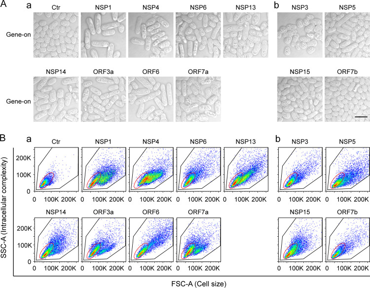
FIG 2.
The effect of SARS-CoV-2 protein on fission yeast cellular morphology. Only those SARS-CoV-2 proteins that affected cell proliferation presented in Fig. 1 are shown here. Complete SARS-CoV-2 genome-wide data on fission yeast cellular morphology are included in Fig. S3. (A) Shows the effect of individual SARS-CoV-2 proteins on fission yeast cell morphology. Each image was taken 48 h agi using bright field microscopy. Scale bar = 10 μM. (B) Overall cell morphology as shown by the forward-scattered analysis. A total of 10,000 cells were measured 48 h agi. The forward-scatter light (FSC) measures the distribution of all cell sizes. The side-scatter light (SSC) determines intracellular complexity. Gene-off, no SARS-CoV-2 protein production; gene-on, SARS-CoV-2 protein produced.

