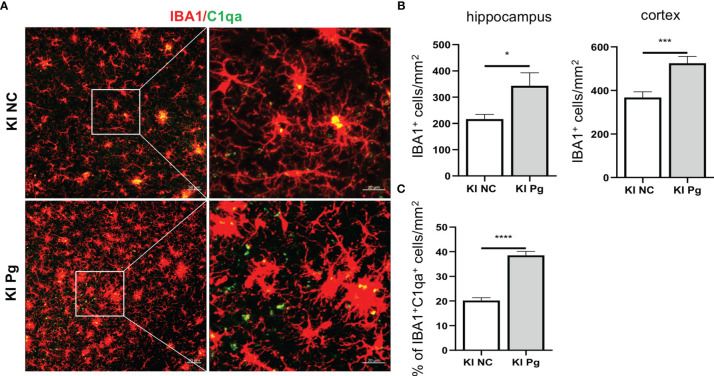
Figure 5.
Pg activates microglia in App KI mice and co-localize with complement C1q. Brain sections from 6-month-old non-infected and Pg-infected App KI mice were immune-stained with antibodies against IBA1and C1qa. (A) Representative Z-stack images of brain sections depicting the spatial association between microglia (red) and C1qa (green). (B) Quantification of IBA1+ cells in the hippocampus and cortex regions. n=6-7 mice/group. (C) Percentage of IBA1+C1qa+ microglia in non-infected and Pg-infected App KI mice. Five representative micrographs of the cortex and hippocampus regions from each mouse were analyzed (n=5-7 mice/group). Data are expressed as mean ± SEM. *P < 0.05, ***P < 0.001, ****P < 0.0001 by unpaired Students t-test.

