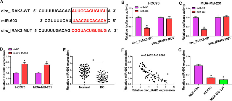
Fig. 3.
Circ_IRAK3 acted as a sponge for miR-603 in BC cells. A The schematic drawing exhibited the putative binding sites between miR-603 and circ_IRAK3. B, C Dual-luciferase reporter assay was conducted to verify the relationship between miR-603 and circ_IRAK3 in HCC70 and MDA-MB-231 cells. D QRT-PCR revealed the expression of miR-603 in HCC70 and MDA-MB-231 cells transfected with si-NC or si-circ_IRAK3. E QRT-PCR exhibited the expression of miR-603 in BC tissues and adjacent non-cancerous tissues. F Pearson’s correlation analysis presented the correlation between miR-603 and circ_IRAK3 in BC tissues. G QRT-PCR presented miR-603 expression in BC cells and the MCF-10A cells. *P < 0.05

