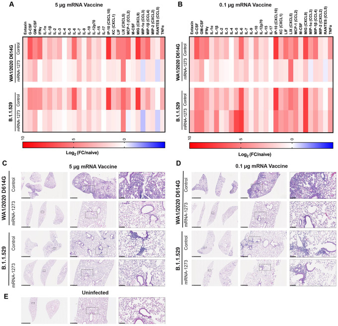
Figure 3. mRNA vaccine protection against disease in K18-hACE2 transgenic mice.
Seven-week-old female K18-hACE2 transgenic mice were immunized with two 5 or 0.1 μg doses of mRNA vaccines, and challenged with WA1/2020 D614G or B.1.1.529. as described in Fig 1 and 2. A-B. Heat-maps of cytokine and chemokine levels in lung homogenates at 6 dpi in animals immunized with 5 μg (A) or 1 μg (B) doses of indicated mRNA vaccines. Fold-change was calculated relative to naive uninfected mice, and log2 values are plotted (2 experiments, n = 7–8 per group except naive, n = 4). The full data set is shown in Table S1–S2. C-E. Hematoxylin and eosin staining of lung sections harvested from control or mRNA-1273 vaccinated animals (5 μg dose, C; 1 μg dose, D) at 6 dpi with WA1/2020 D614G or B.1.1.529. A section from an uninfected animal (E) is shown for comparison. Images show low- (left; scale bars, 1 mm), moderate- (middle, scale bars, 200 μm), and high-power (bottom; scale bars, 50 μm). Representative images of multiple lung sections from n = 3 per group.

