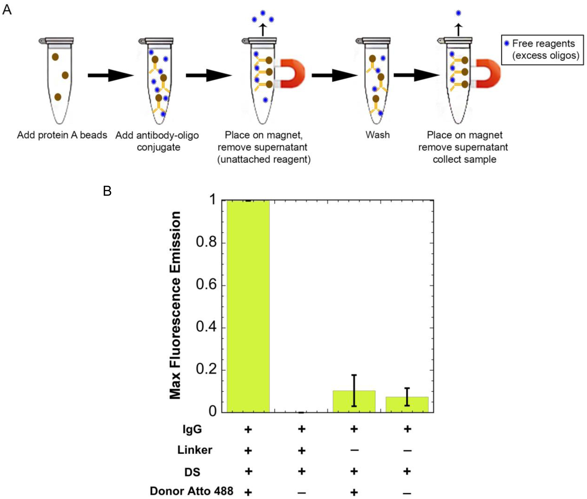
Figure 4:

Separating free reagents using protein A beads. (A) The antibody-oligo conjugate (red Y with a blue circle attached) is added to the protein A beads (brown circle) and is incubated with rotation for 10 minutes. It is then placed on a magnet, pulling the beads out of solution, and the supernatant containing free reagents (unattached blue circles) is removed. The final product is collected containing the antibody-oligo conjugate. (B) Maximum fluorescence intensity values when excited at 450nm. Results show an increased fluorescence signal for the donor when the linker is added. Without the linker, the fluorescence signal for the donor is the same intensity as the background fluorescence.
