
Figure 1.
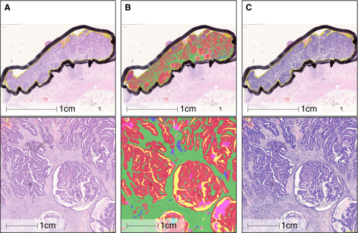
Example of a tumour annotated using a deep neural net algorithm, showing overview and zoomed view. A, Unannotated original; B, deep neural network (DNN) annotation; C, cell segmentation. Tissue compartments: red = epithelium; green = desmoplastic stroma; purple = inflamed stroma; blue = mucin; yellow = white space; pink = necrosis.