Abstract
Objective:
To clinically evaluate the alignment efficiency of 0.016-inch coaxial superelastic nickel-titanium (NiTi) and 0.016-inch superelastic NiTi in the lower anterior region over a period of 12 weeks.
Materials and Methods:
A sample of 24 patients requiring lower anterior alignment were included in this single-center, single-operator, double-blind clinical trial and were randomly allocated into two groups of 12 patients. The type of wire selected for each patient was not disclosed to the provider or to the patient. Comparison of pretreatment characteristics of the archwire groups revealed no discrimination between two samples, thus verifying the random allocation of the intervention. An initial alginate impression of the lower arch was followed by impressions at 4-, 8-, and 12-week intervals. Casts were measured using the coordinate measuring machine to denote the degree of alignment. Duplicate readings of the cast series were taken to assess measurement variation.
Results:
A statistically significant difference (P < .05) in the mean values of tooth movement demonstrated the superior aligning efficiency of coaxial superelastic NiTi over single-stranded superelastic NiTi in relieving lower anterior crowding. The measurement error recorded was within acceptable limits, with range values within 95% limits of agreement.
Conclusion:
Coaxial superelastic NiTi wire proved superior to single-stranded NiTi in its efficiency in relieving lower anterior crowding over a 12-week period.
Keywords: Orthodontic archwires, Superelastic NiTi, Mandibular anterior de-crowding, Coaxial superelastic NiTi, Alignment archwires
INTRODUCTION
In orthodontics, archwires that can deliver light continuous forces over long distances have been shown in vivo to be most efficient in the alignment phase.1 Attempts to reduce the load deflection ratio led to the introduction of multistranded steel wires, nonsuperelastic or martensitic stabilized nickel-titanium (NiTi) wires, superelastic or austenitic active NiTi wires, and true shape memory or martensitic active wires for initial alignment.2 Clinical trials conducted to assess the alignment efficiency of these archwires failed to demonstrate any significant advantage of any particular wire in overall alignment of the arches.3–11
Multistranding of archwires to gain mechanical advantages such as increased flexibility and a reduced load deflection rate has been successfully attempted with the use of stainless steel.12–15 The same concept was applied in the case of superelastic NiTi by Hanson (cited by Berger and Waram16); this led to the introduction of Supercable, a seven-stranded round coaxial superelastic NiTi archwire.16–18 According to manufacturers, this wire will not take a set after bending. Laboratory tests seem to suggest that these wires exert only 36% to 70% of the force of solid NiTi wires.16,17
The aim of the present clinical study was to evaluate whether there is any significant difference in the extent of lower anterior alignment and leveling between 0.016-inch coaxial superelastic NiTi and 0.016-inch single-stranded superelastic NiTi by measuring the amount of tooth movement that occurred at 4-, 8-, and 12-week intervals.
MATERIALS AND METHODS
Archwire specimens in the study included (1) 0.016-inch coaxial superelastic wire (Regular 7 Stranded Supercable Wire, Speed System Orthodontics, Ontario, Canada) and (2) 0.016-inch single-stranded superelastic wire (Rematitan Lite Wire, Dentauram GmbH & Co KG, Ispringen, Germany). On the basis of a previous pilot study, the likely size of differences and measured errors was predicted. For an alpha error of .05 and power of 95%, assuming that the change in measurements at 4 weeks for the Rematitan and Supercable groups was 1.5 mm and 2.5 mm, respectively, with a standard deviation (SD) of 0.60 (based on the pilot study), the minimum sample size required was estimated to be 10 for each of the two groups. It was decided to select 24 subjects to be randomized to two groups of 12, with the expectation of dropouts, but all remained until completion of the trial. Institutional approval was received for this study, as was patient consent.
Inclusion Criteria
Twenty-four patients drawn from a single center and treated under a single operator requiring lower anterior alignment were included in this double-blind investigation. Selection of participants from a large pool of patients was based on the following inclusion criteria:
Female patients in postmenarche period between 13 and 15 years of age with crowding in the lower anterior segment and having a mandibular irregularity index greater than 6
Class I skeletal pattern
Nonextraction treatment in mandibular arch
Eruption of all mandibular teeth with no spacing between them
No relevant medical history
No recent history of intake of drugs such as nonsteroidal anti-inflammatory drugs (NSAIDs)
Patients who may have experienced periodontal disease and hence loss of attachment was avoided
No previous active orthodontic treatment
Full arch mechanics, preadjusted edgewise appliance therapy
No therapeutic intervention planned involving intermaxillary or other intraoral or extraoral appliances during the study period
Participants were told to avoid intake of drugs during the study period. If they had taken any drugs because of unavoidable circumstances, they were requested to report the matter. The intention was to exclude them from the study in such instances. The allocation of archwire was predetermined and randomized. Randomization was done using computer software–generated numbers. Opaque envelopes were used to allocate the archwires to two groups, each consisting of 12 participants. Allocation thus was concealed from the investigator and from participants during the study. All patients were bonded with 0.022 × 0.028-inch slot MBT prescription brackets (Victory Series, 3M Unitek, Monrovia, Calif). Bracket bonding, archwire placement, and treatment were performed by the same clinician.
Data Collection
For each patient, a good-quality alginate impression of each dental arch was taken at the prealignment phase after placement of bonded attachments and bands. The allocated archwire was then ligated as fully as possible into the bracket, usually with elastomeric modules. In cases where it was not possible to engage the archwire with elastomeric module, the tooth was ligated with steel ties. At the routine follow-up appointment at 4 weeks, the wire was removed and another alginate impression was made. This alginate impression was then cast with stone. The archwires were ligated and activated with elastomeric modules or steel ties. The whole procedure was repeated again after 8 and 12 weeks.
Measurement of Study Casts
The models were trimmed for ease of horizontal orientation to the table of the coordinate measuring machine (Figures 1 and 2).19,20 Individual points were noted for each tooth from the mesial aspect of the canine to the contralateral canine in three dimensions and were digitized in turn. For canines, the external and occlusal line angle of the bracket was used; for incisors, the incisal edges were used. No bracket failures were noted during the period of study.
Figure 1.
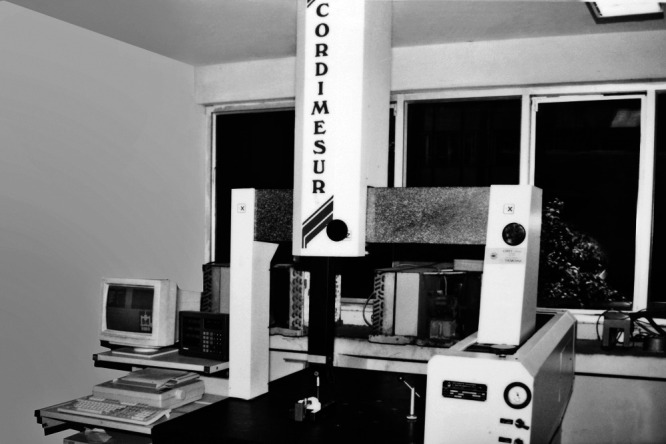
Coordinate measuring machine.
Figure 2.
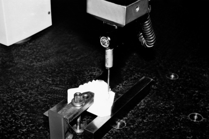
Close view of the coordinate measuring machine measuring the specimen.
Raw data consisted of measurements of each of the five intertooth contact point distances within the lower anterior segment. All readings were measured by an expert single operator, who was not aware of the archwire specimen used for the arches being measured. These readings were available at 0-, 4-, 8-, and 12-week stages. Data for intertooth distances (3-2, 2-1, 1-1, 1-2, 2-3) were collated, and changes at all intertooth distances were summated to represent overall tooth movement, thus deriving a figure for the average alignment of the lower anterior segment at each stage.
Relevant pretreatment characteristics of the two groups were compared using t-tests. Repeated measures analysis of variance (ANOVA) was used to assess the statistical significance of differences in measurements between the two groups, with adjustment for variations in measurements across the three time points. At each time point, mean values of the two groups were compared with the use of Student's t-test. A significance level of P < .05 was used for all tests.
Duplicate readings of the cast series were taken at the commencement and at the end of the trial to assess measurement variation. Intrarater reliability of measurements was assessed from readings taken of the cast series at the commencement and at the end of the trial by determining the reliability SD and 95% confidence intervals.
RESULTS
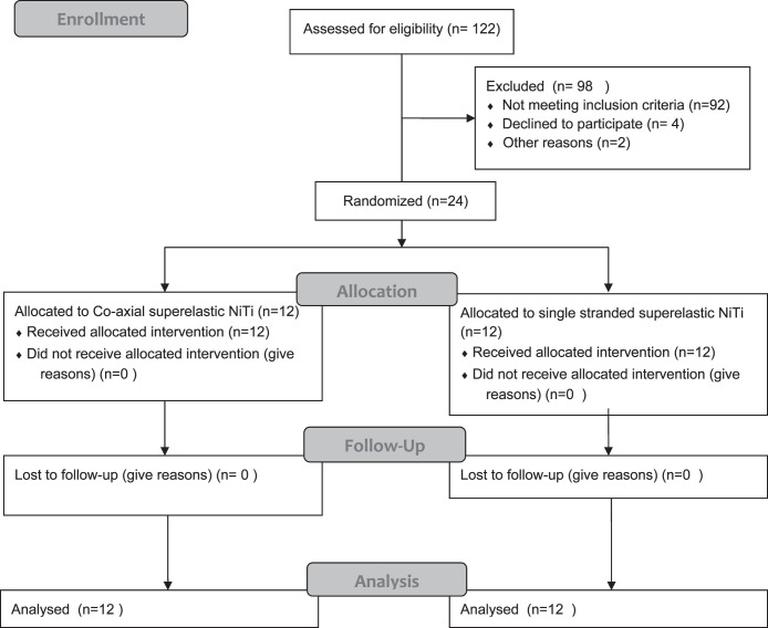
The CONSORT flow diagram for the participation of study subjects is provided as Figure 3. Pretreatment characteristics of the archwire groups are summarized in Table 1. No variable was identified to differentiate the two samples with the use of t-tests, thus verifying the random allocation of interventions to the two wire groups.
Figure 3.
CONSORT flowchart diagram of the clinical trial.
Table 1.
Pretreatment Characteristics of the Archwire Groupsa
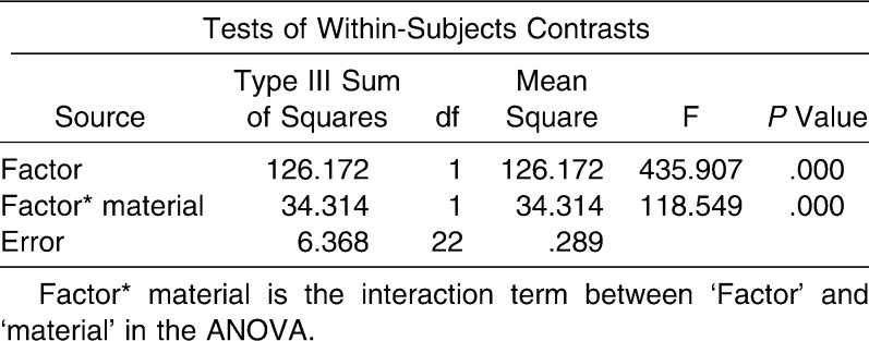
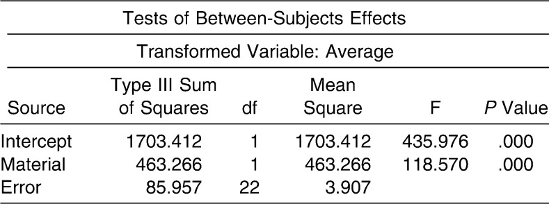
In Tables 2 and 3, repeated measures ANOVA shows the difference between the two materials to be statistically significant (P < .05). For this analysis, repeated measurements were taken as a “factor” and a dichotomous variable “material” to differentiate the two wires. Results inferred that there is a highly statistically significant difference in the mean values of tooth movements (mm) used for two types of wires—Rematitan and Supercable—at 4 weeks', 8 weeks', and 12 weeks' duration. In Table 4, statistical analysis by Student's t-test shows that tooth movement by Supercable wire is significantly greater than mean tooth movement by Rematitan wire at 4 weeks, 8 weeks, and 12 weeks (P < .05).
Table 2.
Results of Repeated Measures Analysis of Variance With Repeated Measurements as “Factor” and a Dichotomous Variable “Material”
Table 3.
Results of Repeated Measures Analysis of Variance With Repeated Measurements as “Factor” and a Dichotomous Variable “Material”
Table 4.
Comparison of Mean Values of Tooth Movement With Two Types of Archwirea
Reproducibility of the measurements was calculated to attain an idea of the measurement error in the study. For this, intertooth distances (3-2, 2-1, 1-1, 1-2, 2-3) were summated for each reading taken for a particular sample. The same was done for the duplicate reading. The difference between these two readings was noted, and the reliability SD was derived.21 Table 5 expresses the reliability SD of the measurements and the 95% limits. Table 5 shows that there is not much measurement error, because the SD of the difference of the two readings observed before and after treatment for both wires is not high. Also, 95% limits of agreement (ie, range values) show consistency between the two readings in all subjects both before and after treatment.
Table 5.
Reproducibility of Measurements
DISCUSSION
According to Proffit,22 and Bennett and McLaughlin,23 the first phase of treatment deals with alignment and correction of vertical and horizontal discrepancies by leveling out the arches, and the initial aligning wires should apply light continuous force. Conventionally, round wires are used for alignment because tightly fitting resilient rectangular archwires produce back-and-forth movement of the root apices as the teeth move into alignment.24 To properly utilize the superelastic properties of NiTi wires, it is necessary to deform them beyond a certain bending angle by as much as 50 to 70 degrees to reach the superelastic plateau.25,26 In clinical practice, such situations are most likely to be seen in the lower anterior region owing to the severity of crowding and the reduced interbracket span. So in this study, the alignment efficiency of 0.016-inch single-stranded superelastic NiTi vs 0.016-inch coaxial superelastic NiTi was compared in severe lower anterior crowding cases.
In our study, instead of taking the mesial and distal anatomic contact points of the teeth as fiducial points, investigators used the external and occlusal line angles of brackets for canines and the incisor edges for incisors to limit point identification error and hence improve point reproducibility.7 The main difference from Little's index27 is that tooth positions were measured in all three dimensions. Here the attempt is made to measure the amount of tooth movement to denote the degree of alignment that has occurred owing to the archwire. So, limitations of Little's index, such as insensitivity to reciprocal rotations and vertical discrepancies of teeth, were avoided.
Results of this study show that the degree of alignment with coaxial superelastic NiTi (Supercable) is greater than with single-stranded superelastic NiTi (Rematitan), and the difference is statistically significant. Because this was a maiden attempt to evaluate the alignment efficiency of coaxial superelastic NiTi at a clinical level, no comparisons could be made. However, results can be compared with those of studies that used NiTi as aligning wire.
Pandis et al.10 found no difference in alleviation of mandibular anterior crowding with copper-nickel-titanium and superelastic NiTi wires. They speculated that this may be due to differences in loading between laboratory conditions and the oral cavity resulting from free play between archwire and bracket slot. Unrealistic force variants at which plateau levels are reached in the stress-strain curve of NiTi wires could be another reason. Evans et al.7 found no statistically significant difference in alignment capabilities of medium force active martensitic rectangular NiTi wire, multistranded stainless steel wire, and graded force active martensitic NiTi wire. However, for studying the alignment capabilities of the anterior segment of the arch, investigators had included upper and lower anterior segments together. The reason why they did not obtain a statistically significant result could be that interbracket distance in the upper anterior segment is greater than in the lower, or that investigators had used rectangular archwires for comparison in the study. West et al.5 compared superelastic NiTi and multistranded stainless steel and reported better alignment with superelastic NiTi in the lower anterior region; this is statistically significant. In their study, the adjusted geometric mean ratio attained for wires over a 6-week period was 1.20 for the lower anterior segment. In our study, mean tooth movement of 1.55 over a 4-week period for superelastic NiTi was noted. Any slight variation may have been due to the difference in crowding in the sample size, or to the method of ligation or inconsistent force levels among different manufacturers or even between different batches of the same manufacturer. Jones et al.4 found greater mean improvement in incisal alignment with superelastic NiTi when compared with coaxial stainless steel even though the improvement was not statistically significant. All these studies point in the direction that lower anterior alignment tends to be better and faster with superelastic NiTi wires than with coaxial stainless steel.
In addition to being an NiTi wire, Supercable provides the advantages of greater springback, increased resistance to deformation, and low force delivery, probably because it is multistranded.16 Another factor that we have to consider is the play between the bracket and the archwire. Proffit24 advises that for mesiodistal sliding along an archwire, at least 2 mil clearance between the archwire and the bracket is needed, and 4 mil clearances are desired. The main difference between the two archwires in our study—coaxial superelastic NiTi and superelastic NiTi—is that although we are engaging archwires of the same diameter, the elastic modulus of the wires is different. So we are able to engage the archwire into the bracket better with coaxial superelastic wire. Attempts to deform the superelastic wire to reach the superelastic plateau by selecting severe lower anterior crowding cases have led to many instances of difficulty in engaging the single-stranded superelastic NiTi and of partial ligation with steel ties. Thus superelastic coaxial wires offer the advantage of engaging a relatively large archwire at the start of treatment with low force delivery. So a greater degree of uprighting, leveling, and rotational control is achieved than with other initial wires. This was perhaps one of the reasons why the difference in alignment of teeth between the archwires was so significant. The other option would be to use a single-stranded superelastic NiTi wire of lesser dimension, such as 0.014 inch, in lower anterior crowding cases at the cost of losing better uprighting, leveling, and rotational control and, in effect, alignment capability as attained with a 0.016-inch dimension wire.
In our study, measurement error calculated for pretreatment samples had a reliability SD value of 0.0127 for Rematitan; for Supercable, it was 0.0579. For posttreatment samples, the reliability SD was 0.0216 for Rematitan and 0.0202 for Supercable. So the measurement error in this study was within acceptable limits, given the mean measurements for tooth movement. Further comparative study of these archwires in cases of less severe crowding and over a longer time is recommended to evaluate the overall effectiveness of coaxial superelastic NiTi in shortening the duration of lower anterior alignment.
CONCLUSION
Superelastic coaxial NiTi wires exhibit a statistically significant difference over superelastic single-stranded NiTi in their alignment efficiency to relieve lower anterior crowding after 4, 8, and 12 weeks.
REFERENCES
- 1.Dalstra M, Melsen B. Does the transition temperature of Cu-NiTi archwires affect the amount of tooth movement during alignment. Orthod Craniofac Res. 2004;7:21–25. doi: 10.1046/j.1601-6335.2003.00275.x. [DOI] [PubMed] [Google Scholar]
- 2.Brantley W. A. Orthodontic wires. In: Brantley W. A, Eliades T, editors. Orthodontic Materials Scientific and Clinical Aspects. Stuttgart, Germany: Thieme; 2001. pp. 91–99. [Google Scholar]
- 3.O'Brien K, Lewis D, Shaw W, Combe E. A clinical trial of aligning archwires. Eur J Orthod. 1990;12:380–384. doi: 10.1093/ejo/12.4.380. [DOI] [PubMed] [Google Scholar]
- 4.Jones M. L, Staniford H, Chan C. Comparison of superelastic NiTi and multistranded stainless steel wires in initial alignment. J Clin Orthod. 1990;24:611–613. [PubMed] [Google Scholar]
- 5.West A. E, Jones M. L, Newcombe R. G. Multiflex versus superelastic: a randomized clinical trial of the tooth alignment ability of initial archwires. Am J Orthod Dentofacial Orthop. 1995;108:464–471. doi: 10.1016/s0889-5406(95)70046-3. [DOI] [PubMed] [Google Scholar]
- 6.Cobb N. W, Kula K. S, Phillips C, Proffit W. R. Efficiency of multi-strand steel, superelastic Ni-Ti and ion-implanted Ni-Ti archwires for initial alignment. Clin Orthod Res. 1998;1:12–19. doi: 10.1111/ocr.1998.1.1.12. [DOI] [PubMed] [Google Scholar]
- 7.Evans T. J, Jones M. L, Newcombe R. G. Clinical comparison and performance perspective of three aligning archwires. Am J Orthod Dentofacial Orthop. 1998;114:32–39. doi: 10.1016/s0889-5406(98)70234-3. [DOI] [PubMed] [Google Scholar]
- 8.Mandall N, Lowe C, Worthington H, et al. Which orthodontic archwire sequence? A randomized clinical trial. Eur J Orthod. 2006;28:561–566. doi: 10.1093/ejo/cjl030. [DOI] [PubMed] [Google Scholar]
- 9.Riley M, Bearn D. R. A systemic review of clinical trials of aligning archwires. J Orthod. 2009;36:42–51. doi: 10.1179/14653120722914. [DOI] [PubMed] [Google Scholar]
- 10.Pandis N, Polychronopoulou A, Eliades T. Alleviation of mandibular anterior crowding with copper-nickel-titanium vs nickel-titanium wires: a double-blind randomized control trial. Am J Orthod Dentofacial Orthop. 2009;136:151–157. doi: 10.1016/j.ajodo.2009.03.030. [DOI] [PubMed] [Google Scholar]
- 11.Ong E, Ho C, Miles P. Alignment efficiency and discomfort of three orthodontic archwire sequences: a randomized clinical trial. J Orthod. 2011;38:32–39. doi: 10.1179/14653121141218. [DOI] [PubMed] [Google Scholar]
- 12.Kusy R. P, Dilley G. J. Elastic modulus of a triple-stranded stainless steel archwire via three- and four-point bending. J Dent Res. 1984;36:1232–1240. doi: 10.1177/00220345840630101401. [DOI] [PubMed] [Google Scholar]
- 13.Kusy R. P, Stevens L. E. Triple-stranded stainless steel wires: evaluation of mechanical properties and comparison with titanium alloy alternatives. Angle Orthod. 1987;57:18–32. doi: 10.1043/0003-3219(1987)057<0018:TSSW>2.0.CO;2. [DOI] [PubMed] [Google Scholar]
- 14.Rucker B. K, Kusy R. P. Theoretical investigation of elastic flexural properties for multistranded orthodontic archwires. J Biomed Mater Res. 2002;62:338–349. doi: 10.1002/jbm.10325. [DOI] [PubMed] [Google Scholar]
- 15.Rucker B. K, Kusy R. P. Elastic flexural properties of multistranded stainless steel versus conventional nickel titanium archwires. Angle Orthod. 2002;72:302–309. doi: 10.1043/0003-3219(2002)072<0302:EFPOMS>2.0.CO;2. [DOI] [PubMed] [Google Scholar]
- 16.Berger J, Waram T. Force levels of nickel titanium initial archwires. J Clin Orthod. 2007;41:286–292. [PubMed] [Google Scholar]
- 17.Berger J, Byloff F. K, Waram T. Supercable and the speed system. J Clin Orthod. 1998;32:246–253. [PubMed] [Google Scholar]
- 18.Speed Supercable Speed System World Wide Website Product Information Catalogue.
- 19.Lundgren D, Moll O, Kurol J, Martensson B. Accuracy of orthodontic force and tooth movement measurements. Br J Orthod. 1996;23:241–248. doi: 10.1179/bjo.23.3.241. [DOI] [PubMed] [Google Scholar]
- 20.Braun S, Hnat W. P, Leschinsky R, Legan H. L. An evaluation of the shape of some popular nickel titanium alloy preformed archwires. Am J Orthod Dentofacial Orthop. 1999;116:1–12. doi: 10.1016/S0889-5406(99)70297-0. [DOI] [PubMed] [Google Scholar]
- 21.Hopkins W. G. A new view of statistics: measures of reliability, 2009. Available at: http://www.sportsci.org/resource/stats/index.html Accessed January 30, 2011. [Google Scholar]
- 22.Proffit W. R, Fields H. W, Sarver D. M. Contemporary Orthodontics 4th ed. St Louis, Mo: Mosby; 2007. pp. 551–552. [Google Scholar]
- 23.Bennett J. C, McLaughlin R. P. Orthodontic Treatment Mechanics and the Preadjusted Appliance 1st ed. Vol. 89 Prescott, Ariz: Wolfe Publishing; 1993. [Google Scholar]
- 24.Proffit W. R, Fields H. W, Sarver D. M. Contemporary Orthodontics 4th ed. Vol. 553 St Louis, Mo: Mosby; 2007. [Google Scholar]
- 25.Meling T. R, Odegaard J. The effect of short-term temperature changes on the mechanical properties of rectangular nickel titanium archwires tested in torsion. Angle Orthod. 1998;68:369–376. doi: 10.1043/0003-3219(1998)068<0369:TEOSTT>2.3.CO;2. [DOI] [PubMed] [Google Scholar]
- 26.Schumacher H. A, Bourauel C, Drescher D. The deactivation behavior and effectiveness of different orthodontic leveling arches—a dynamic analysis of the force. Fortschr Kieferorthop. 1992;53:273–285. doi: 10.1007/BF02325076. [DOI] [PubMed] [Google Scholar]
- 27.Little R. M. The irregularity index: a quantitative score of mandibular anterior alignment. Am J Orthod. 1975;68:554–563. doi: 10.1016/0002-9416(75)90086-x. [DOI] [PubMed] [Google Scholar]


