Figure 1.
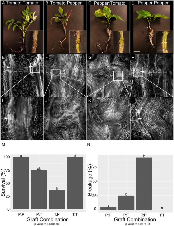
Heterografted tomato and pepper plants show severe vascular patterning defects, reduced viability, and biomechanical failure 30 DAG. A–D, Representative images of self-grafted tomato (A), heterografted tomato:pepper (B), pepper:tomato (C), and self-grafted pepper (D) plants taken 30 DAG. White arrows indicate graft junctions. E–L, High-resolution confocal imaging of vascular anatomy for self-grafted tomato (E and I), heterografted tomato:pepper (F and J), and pepper:tomato (G and K), and self-grafted pepper (H and L) plants taken at 30 DAG. Tissues were stained with PI to visualize cell walls, and cleared in methyl salicylate. White arrowheads point to xylem bridges. Dashed lines represent the graft site. (M and N) Heterografts exhibited significantly reduced viability relative to self-grafted plants (M), and higher breakage along the graft site during bend tests (N). P-values under graphs shown from Fisher’s exact test. Different letters indicate significant differences in the graft combinations (pairwise comparisons using Fisher’s exact test, P < 0.05, P-values shown in Supplemental Data Set 1). P:P = pepper:pepper graft, T:T = tomato:tomato graft, P:T = pepper:tomato graft, T:P = tomato:pepper graft, PI = Propidium Iodide. N = 3 (A–L), n = 12–18 (M, N). Scale bars = 2 cm (A–D), 1 cm (E–H), 400 µm (I–L).

