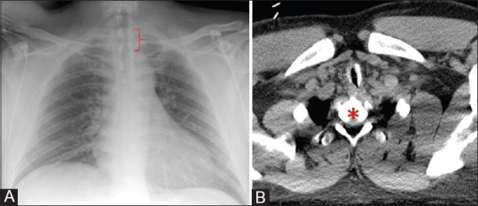
Figure 1.

Panel A – Upright chest x-ray demonstrating stenotic tracheal segment measuring 7.5 mm diameter by 3.3 cm length (bracket). Panel B - Computed tomography demonstrating tracheal stenosis narrowest transverse section of 7.5 mm (arrow) at the level of first thoracic vertebral level (asterisk)
