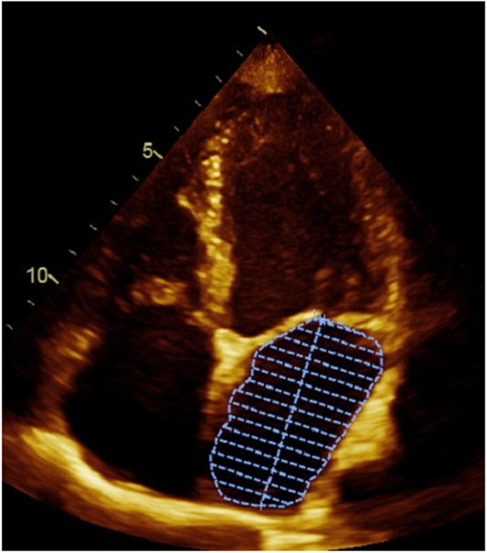
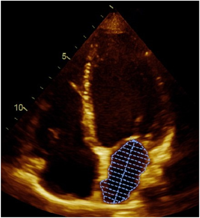
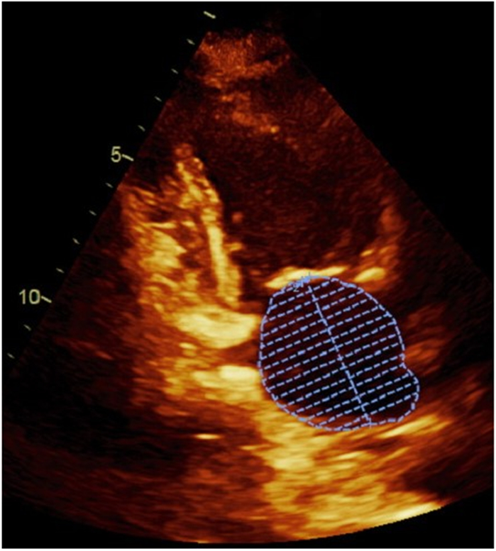
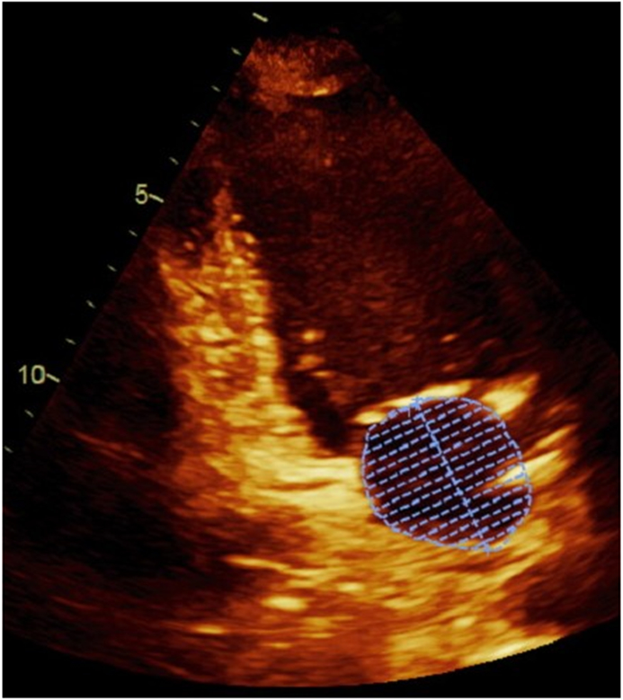
Figure 1. Left atrial volume measurements in apical 4-chamber and 2-chamber views.


Panel A demonstrates the measurement of the largest left atrial volume at end-systole (LAESVI) while panel B demonstrates the measurement of the smallest left atrial volume at end-diastole (LAEDVI) in the apical 4-chamber view. Panel C demonstrates the measurement of LAESVI and panel D demonstrates the measurement of LAEDVI in the apical 2-chamber view.
