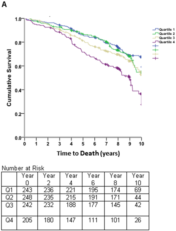
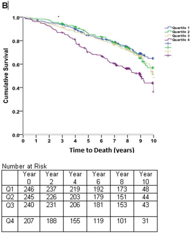
Figure 4. Survival Free of Death.


Kaplan-Meier curves of time to death in subjects with stable coronary heart disease and preserved left ventricular systolic function, stratified by quartiles of the left atrial end-diastolic volume index (LAEDVI; Panel A) and left atrial volume index (LAESVI; Panel B). The rate of death was highest in subjects in the highest quartile and lowest in subjects in the lowest quartile. The separation of curves occurred earlier for LAEDVI compared to LAESVI.
