. 2022 Jan 24;11:e69094. doi: 10.7554/eLife.69094

Figure 2. Through-cuticle imaging of the fly brain with 2P and 3P excitation.

(A) Schematic of the multiphoton microscope setup. Fly head is fixed to a cover slip and placed under the objective (HWP, half-wave plate; PBS, polarization beam splitter; PMT, photomultiplier tube). The imaging window on the fly head is shown in the picture (lower left). Scale bar = 200 µm. (B–D) The head-uncompressed and head-compressed imaging preparations. The first column shows the side image of the fly that is head fixed to the cover glass (scale bar = 1 mm). The second and third columns show the fly head visualized under a brightfield (top view) and fluorescent dissecting microscopes (widefield-fluo), respectively. Arrows and the rectangle area in widefield-fluo column indicate the imaging window (scale bar = 200 µm). (E–G) Cross-section imaging of the mushroom body Kenyon cells expressing CD8-GFP through the head cuticle at 920 nm (2P) and 1320 nm (3P) excitation. The Z projections of 2P (cyan, left) and 3P (green, right) imaging stacks. For each imaging preparation, the same fly head is imaged with 3P and 2P excitation (scale bar = 20 µm).

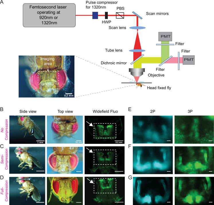
Figure 2.

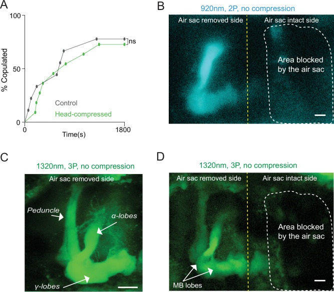
Figure 2—figure supplement 1. Head compression does not affect male courtship and removal of air sacs allows 2P and 3P imaging in a head-uncompressed preparation.

Figure 2—figure supplement 1.

(A) Male copulation percentage is quantified in controls and flies that were previously head compressed (n = 7–8, log-rank (Mantel-Cox) test, p>0.05). (B–D) Cross-section imaging of the mushroom body Kenyon cells expressing GCaMP6s through the head cuticle at 920 nm (2P) and 1320 nm (3P) excitation after removing air sacs on one side of the head. The fly head is not compressed to the cover slip. The Z projections of (B) 2P (cyan) and (C, D) 3P (green) imaging stacks. For each imaging preparation, the same fly brain is imaged with 3P and 2P excitation. Mushroom body structures are visible on the side where air sacs are removed. The dotted lines show the area where the air sacs block imaging. (C) Zoomed-in 3P image taken from the side of the brain where the air sacs are removed. Mushroom body lobes and the peduncle are clearly visible in the air sac removed side without head compression (scale bars = 20 µm).