Figure 2.

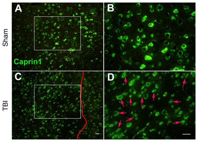
Moderate TBI induces the alteration of caprin1 expression in injured neurons of the C577BL/6J mouse motor cortex. (A and B) Sham group at magnifications of (A) ×100 and (B) ×200. (C and D) TBI group at (C) ×100 and (D) ×200 magnification (scale bars, 30 µm). B and D provide magnified windows of A and C, respectively. In TBI mice, the right somatosensory cortex had been subjected to a moderate fluid percussion pulse from 2.5 to 2.6 atm. On day 8 post-injury, immunofluorescence analysis indicated that neurons in the sham animals exhibited clear and even caprin1 expression in the motor cortex; while the expression of caprin1 (stress granule-related RNA-binding proteins, as indicated by red arrows in D) in typical lesioned neurons on the left of the vertical red line in TBI animals was largely diffusive and uneven with numerous dots. However, caprin1 expression in neurons outside the lesion zone (on the right side of the vertical red line) was less affected. The experimental protocol is provided in Appendix S1. TBI, traumatic brain injury.
